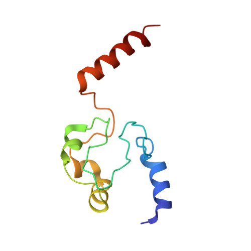
3NBT

Crystal structure of trimeric cytochrome c from horse heart


Experimental Data Snapshot

  • Method: X-RAY DIFFRACTION
  • Resolution: 2.10 Å
  • R-Value Free: 0.254 
  • R-Value Work: 0.211 
  • R-Value Observed: 0.211 

Starting Model: experimental
View more details

wwPDB Validation   3D Report Full Report


Ligand Structure Quality Assessment 


This is version 2.2 of the entry. See complete history


Literature

Cytochrome c polymerization by successive domain swapping at the C-terminal helix

Hirota, S.Hattori, Y.Nagao, S.Taketa, M.Komori, H.Kamikubo, H.Wang, Z.Takahashi, I.Negi, S.Sugiura, Y.Kataoka, M.Higuchi, Y.

(2010) Proc Natl Acad Sci U S A 107: 12854-12859

  • DOI: https://doi.org/10.1073/pnas.1001839107
  • Primary Citation of Related Structures:  
    3NBS, 3NBT

  • PubMed Abstract: 

    Cytochrome c (cyt c) is a stable protein that functions in a monomeric state as an electron donor for cytochrome c oxidase. It is also released to the cytosol when permeabilization of the mitochondrial outer membrane occurs at the early stage of apoptosis. For nearly half a century, it has been known that cyt c forms polymers, but the polymerization mechanism remains unknown. We found that cyt c forms polymers by successive domain swapping, where the C-terminal helix is displaced from its original position in the monomer and Met-heme coordination is perturbed significantly. In the crystal structures of dimeric and trimeric cyt c, the C-terminal helices are replaced by the corresponding domain of other cyt c molecules and Met80 is dissociated from the heme. The solution structures of dimeric, trimeric, and tetrameric cyt c were linear based on small-angle X-ray scattering measurements, where the trimeric linear structure shifted toward the cyclic structure by addition of PEG and (NH(4))(2)HPO(4). The absorption and CD spectra of high-order oligomers (approximately 40 mer) were similar to those of dimeric and trimeric cyt c but different from those of monomeric cyt c. For dimeric, trimeric, and tetrameric cyt c, the DeltaH of the oligomer dissociation to monomers was estimated to be about -20 kcal/mol per protomer unit, where Met-heme coordination appears to contribute largely to DeltaH. The present results suggest that cyt c polymerization occurs by successive domain swapping, which may be a common mechanism of protein polymerization.


  • Organizational Affiliation

    Graduate School of Materials Science, Nara Institute of Science and Technology, 8916-5 Takayama, Ikoma, Nara 630-0192, Japan. hirota@ms.naist.jp


Macromolecules
Find similar proteins by:  (by identity cutoff)  |  3D Structure
Entity ID: 1
MoleculeChains Sequence LengthOrganismDetailsImage
Cytochrome c
A, B, C, D, E
A, B, C, D, E, F
104Equus caballusMutation(s): 0 
UniProt
Find proteins for P00004 (Equus caballus)
Explore P00004 
Go to UniProtKB:  P00004
Entity Groups  
Sequence Clusters30% Identity50% Identity70% Identity90% Identity95% Identity100% Identity
UniProt GroupP00004
Sequence Annotations
Expand
  • Reference Sequence
Small Molecules
Ligands 4 Unique
IDChains Name / Formula / InChI Key2D Diagram3D Interactions
HEC
Query on HEC

Download Ideal Coordinates CCD File 
G [auth A]
J [auth B]
M [auth C]
P [auth D]
S [auth E]
G [auth A],
J [auth B],
M [auth C],
P [auth D],
S [auth E],
V [auth F]
HEME C
C34 H34 Fe N4 O4
HXQIYSLZKNYNMH-LJNAALQVSA-N
PG4
Query on PG4

Download Ideal Coordinates CCD File 
H [auth A]
K [auth B]
N [auth C]
Q [auth D]
T [auth E]
H [auth A],
K [auth B],
N [auth C],
Q [auth D],
T [auth E],
W [auth F]
TETRAETHYLENE GLYCOL
C8 H18 O5
UWHCKJMYHZGTIT-UHFFFAOYSA-N
PGE
Query on PGE

Download Ideal Coordinates CCD File 
R [auth D],
X [auth F]
TRIETHYLENE GLYCOL
C6 H14 O4
ZIBGPFATKBEMQZ-UHFFFAOYSA-N
PEG
Query on PEG

Download Ideal Coordinates CCD File 
I [auth A],
L [auth B],
O [auth C],
U [auth E]
DI(HYDROXYETHYL)ETHER
C4 H10 O3
MTHSVFCYNBDYFN-UHFFFAOYSA-N
Experimental Data & Validation

Experimental Data

  • Method: X-RAY DIFFRACTION
  • Resolution: 2.10 Å
  • R-Value Free: 0.254 
  • R-Value Work: 0.211 
  • R-Value Observed: 0.211 
  • Space Group: P 21 21 21
Unit Cell:
Length ( Å )Angle ( ˚ )
a = 57.204α = 90
b = 95.661β = 90
c = 130.935γ = 90
Software Package:
Software NamePurpose
HKL-2000data collection
MOLREPphasing
CNSrefinement
HKL-2000data reduction
HKL-2000data scaling

Structure Validation

View Full Validation Report


Ligand Structure Quality Assessment 


Entry History 

Deposition Data

Revision History  (Full details and data files)

  • Version 1.0: 2010-07-14
    Type: Initial release
  • Version 1.1: 2011-07-13
    Changes: Version format compliance
  • Version 1.2: 2017-11-08
    Changes: Refinement description
  • Version 2.0: 2019-10-02
    Changes: Advisory, Atomic model, Data collection, Derived calculations, Non-polymer description, Structure summary
  • Version 2.1: 2023-11-01
    Changes: Data collection, Database references, Derived calculations, Refinement description
  • Version 2.2: 2024-11-06
    Changes: Structure summary