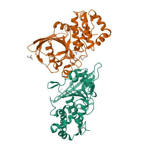
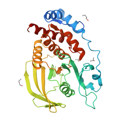
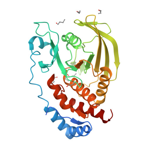
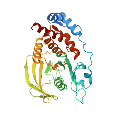
3S3E

Crystal structure of the catalytic domain of PTP10D from Drosophila melanogaster


Experimental Data Snapshot

  • Method: X-RAY DIFFRACTION
  • Resolution: 2.40 Å
  • R-Value Free: 0.227 
  • R-Value Work: 0.205 
  • R-Value Observed: 0.206 

Starting Model: experimental
View more details

wwPDB Validation   3D Report Full Report


This is version 1.2 of the entry. See complete history


Literature

Conformational basis for substrate recruitment in protein tyrosine phosphatase 10D

Madan, L.L.Gopal, B.

(2011) Biochemistry 50: 10114-10125

  • DOI: https://doi.org/10.1021/bi201092q
  • Primary Citation of Related Structures:  
    3S3E, 3S3F, 3S3H, 3S3K

  • PubMed Abstract: 

    The coordinated activity of protein tyrosine phosphatases (PTPs) is crucial for the initiation, modulation, and termination of diverse cellular processes. The catalytic activity of this protein depends on a nucleophilic cysteine at the active site that mediates the hydrolysis of the incoming phosphotyrosine substrate. While the role of conserved residues in the catalytic mechanism of PTPs has been extensively examined, the diversity in the mechanisms of substrate recognition and modulation of catalytic activity suggests that other, less conserved sequence and structural features could contribute to this process. Here we describe the crystal structures of Drosophila melanogaster PTP10D in the apo form as well as in a complex with a substrate peptide and an inhibitor. These studies reveal the role of aromatic ring stacking interactions at the boundary of the active site of PTPs in mediating substrate recruitment. We note that phenylalanine 76, of the so-called KNRY loop, is crucial for orienting the phosphotyrosine residue toward the nucleophilic cysteine. Mutation of phenylalanine 76 to leucine results in a 60-fold decrease in the catalytic efficiency of the enzyme. Fluorescence measurements with a competitive inhibitor, p-nitrocatechol sulfate, suggest that Phe76 also influences the formation of the enzyme-substrate intermediate. The structural and biochemical data for PTP10D thus highlight the role of relatively less conserved residues in PTP domains in both substrate recruitment and modulation of reaction kinetics.


  • Organizational Affiliation

    Molecular Biophysics Unit, Indian Institute of Science, Bangalore 560 012, India.


Macromolecules
Find similar proteins by:  (by identity cutoff)  |  3D Structure
Entity ID: 1
MoleculeChains Sequence LengthOrganismDetailsImage
Tyrosine-protein phosphatase 10D
A, B
307Drosophila melanogasterMutation(s): 0 
Gene Names: CG1817Ptp10D
EC: 3.1.3.48
UniProt
Find proteins for P35992 (Drosophila melanogaster)
Explore P35992 
Go to UniProtKB:  P35992
Entity Groups  
Sequence Clusters30% Identity50% Identity70% Identity90% Identity95% Identity100% Identity
UniProt GroupP35992
Sequence Annotations
Expand
  • Reference Sequence
Small Molecules
Ligands 3 Unique
IDChains Name / Formula / InChI Key2D Diagram3D Interactions
BU1
Query on BU1

Download Ideal Coordinates CCD File 
G [auth B]1,4-BUTANEDIOL
C4 H10 O2
WERYXYBDKMZEQL-UHFFFAOYSA-N
1BO
Query on 1BO

Download Ideal Coordinates CCD File 
D [auth A]
F [auth A]
H [auth B]
I [auth B]
J [auth B]
D [auth A],
F [auth A],
H [auth B],
I [auth B],
J [auth B],
K [auth B],
M [auth B]
1-BUTANOL
C4 H10 O
LRHPLDYGYMQRHN-UHFFFAOYSA-N
IPA
Query on IPA

Download Ideal Coordinates CCD File 
C [auth A],
E [auth A],
L [auth B]
ISOPROPYL ALCOHOL
C3 H8 O
KFZMGEQAYNKOFK-UHFFFAOYSA-N
Experimental Data & Validation

Experimental Data

  • Method: X-RAY DIFFRACTION
  • Resolution: 2.40 Å
  • R-Value Free: 0.227 
  • R-Value Work: 0.205 
  • R-Value Observed: 0.206 
  • Space Group: P 31 2 1
Unit Cell:
Length ( Å )Angle ( ˚ )
a = 102.46α = 90
b = 102.46β = 90
c = 171.842γ = 120
Software Package:
Software NamePurpose
HKL-2000data collection
PHASERphasing
REFMACrefinement
MOSFLMdata reduction
SCALAdata scaling

Structure Validation

View Full Validation Report


Entry History 

Deposition Data

Revision History  (Full details and data files)

  • Version 1.0: 2011-11-02
    Type: Initial release
  • Version 1.1: 2013-07-03
    Changes: Database references
  • Version 1.2: 2023-11-01
    Changes: Data collection, Database references, Derived calculations, Refinement description