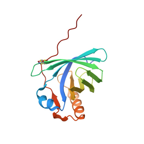
3SAO

The Siderocalin Ex-FABP functions through dual ligand specificities


Experimental Data Snapshot

  • Method: X-RAY DIFFRACTION
  • Resolution: 1.80 Å
  • R-Value Free: 0.233 
  • R-Value Work: 0.202 
  • R-Value Observed: 0.204 

Starting Models: experimental
View more details

wwPDB Validation   3D Report Full Report


Ligand Structure Quality Assessment 


This is version 1.1 of the entry. See complete history


Literature

Galline Ex-FABP Is an Antibacterial Siderocalin and a Lysophosphatidic Acid Sensor Functioning through Dual Ligand Specificities.

Correnti, C.Clifton, M.C.Abergel, R.J.Allred, B.Hoette, T.M.Ruiz, M.Cancedda, R.Raymond, K.N.Descalzi, F.Strong, R.K.

(2011) Structure 19: 1796-1806

  • DOI: https://doi.org/10.1016/j.str.2011.09.019
  • Primary Citation of Related Structures:  
    3SAO

  • PubMed Abstract: 

    Galline Ex-FABP was identified as another candidate antibacterial, catecholate siderophore binding lipocalin (siderocalin) based on structural parallels with the family archetype, mammalian Siderocalin. Binding assays show that Ex-FABP retains iron in a siderophore-dependent manner in both hypertrophic and dedifferentiated chondrocytes, where Ex-FABP expression is induced after treatment with proinflammatory agents, and specifically binds ferric complexes of enterobactin, parabactin, bacillibactin and, unexpectedly, monoglucosylated enterobactin, which does not bind to Siderocalin. Growth arrest assays functionally confirm the bacteriostatic effect of Ex-FABP in vitro under iron-limiting conditions. The 1.8 Å crystal structure of Ex-FABP explains the expanded specificity, but also surprisingly reveals an extended, multi-chambered cavity extending through the protein and encompassing two separate ligand specificities, one for bacterial siderophores (as in Siderocalin) at one end and one specifically binding copurified lysophosphatidic acid, a potent cell signaling molecule, at the other end, suggesting Ex-FABP employs dual functionalities to explain its diverse endogenous activities.


  • Organizational Affiliation

    Division of Basic Sciences, Fred Hutchinson Cancer Research Center, Seattle, WA 98109, USA.


Macromolecules
Find similar proteins by:  (by identity cutoff)  |  3D Structure
Entity ID: 1
MoleculeChains Sequence LengthOrganismDetailsImage
Extracellular fatty acid-binding protein
A, B
160Gallus gallusMutation(s): 3 
Gene Names: EXFABP
UniProt
Find proteins for P21760 (Gallus gallus)
Explore P21760 
Go to UniProtKB:  P21760
Entity Groups  
Sequence Clusters30% Identity50% Identity70% Identity90% Identity95% Identity100% Identity
UniProt GroupP21760
Sequence Annotations
Expand
  • Reference Sequence
Small Molecules
Ligands 4 Unique
IDChains Name / Formula / InChI Key2D Diagram3D Interactions
NKN
Query on NKN

Download Ideal Coordinates CCD File 
C [auth A],
D [auth B]
(2R)-2-hydroxy-3-(phosphonooxy)propyl tetradecanoate
C17 H35 O7 P
FAZBDRGXCKPVJU-MRXNPFEDSA-N
DBH
Query on DBH

Download Ideal Coordinates CCD File 
E [auth B]2,3-DIHYDROXY-BENZOIC ACID
C7 H6 O4
GLDQAMYCGOIJDV-UHFFFAOYSA-N
GOL
Query on GOL

Download Ideal Coordinates CCD File 
G [auth B]GLYCEROL
C3 H8 O3
PEDCQBHIVMGVHV-UHFFFAOYSA-N
FE
Query on FE

Download Ideal Coordinates CCD File 
F [auth B]FE (III) ION
Fe
VTLYFUHAOXGGBS-UHFFFAOYSA-N
Experimental Data & Validation

Experimental Data

  • Method: X-RAY DIFFRACTION
  • Resolution: 1.80 Å
  • R-Value Free: 0.233 
  • R-Value Work: 0.202 
  • R-Value Observed: 0.204 
  • Space Group: P 21 21 21
Unit Cell:
Length ( Å )Angle ( ˚ )
a = 37.95α = 90
b = 68.89β = 90
c = 117.4γ = 90
Software Package:
Software NamePurpose
d*TREKdata reduction
REFMACrefinement
PDB_EXTRACTdata extraction
ADSCdata collection
d*TREKdata scaling
PHASERphasing

Structure Validation

View Full Validation Report


Ligand Structure Quality Assessment 


Entry History 

Deposition Data

Revision History  (Full details and data files)

  • Version 1.0: 2011-12-28
    Type: Initial release
  • Version 1.1: 2023-09-13
    Changes: Data collection, Database references, Derived calculations, Refinement description, Structure summary