3SJK

Crystal structure of the C147A mutant 3C from enterovirus 71


Experimental Data Snapshot

  • Method: X-RAY DIFFRACTION
  • Resolution: 2.10 Å
  • R-Value Free: 0.238 
  • R-Value Work: 0.207 
  • R-Value Observed: 0.209 

Starting Model: experimental
View more details

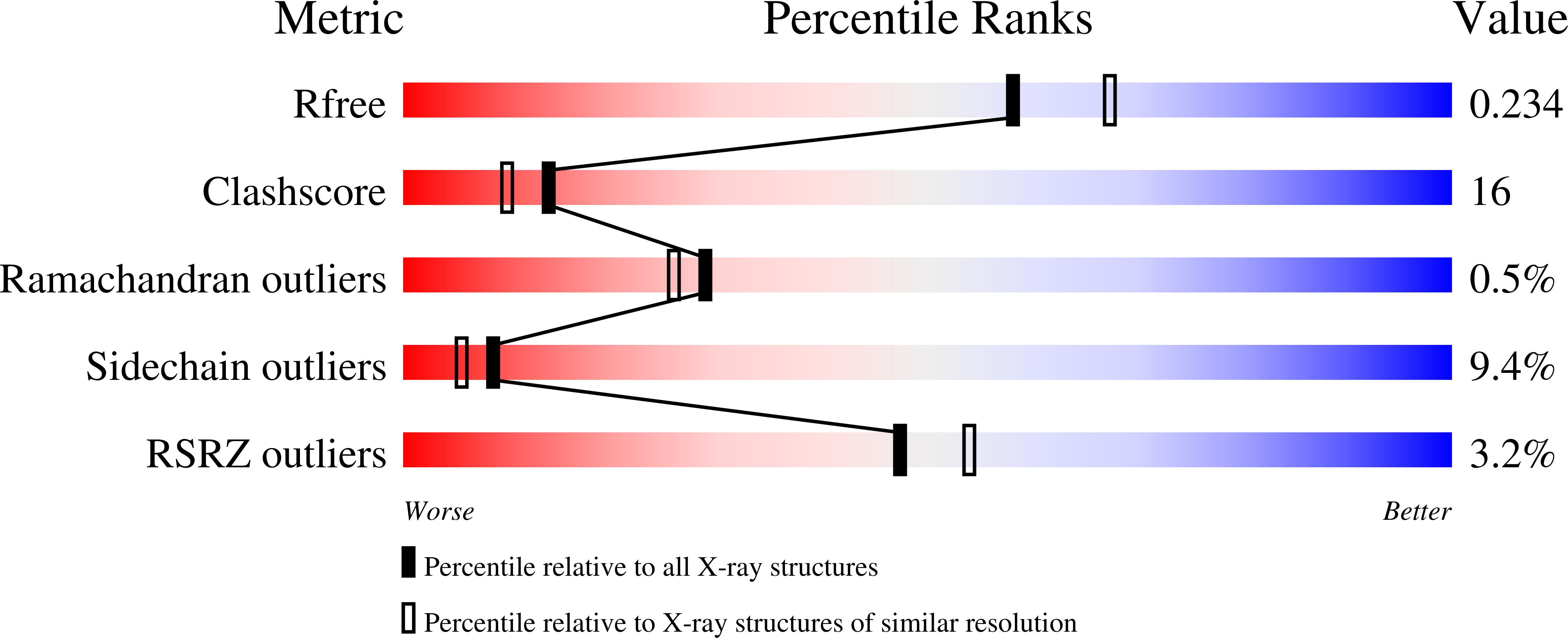
wwPDB Validation   3D Report Full Report


This is version 1.2 of the entry. See complete history


Literature

Enterovirus 71 and Coxsackievirus A16 3C Proteases: Binding to Rupintrivir and Their Substrates and Anti-Hand, Foot, and Mouth Disease Virus Drug Design.

Lu, G.Qi, J.Chen, Z.Xu, X.Gao, F.Lin, D.Qian, W.Liu, H.Jiang, H.Yan, J.Gao, G.F.

(2011) J Virol 85: 10319-10331

  • DOI: https://doi.org/10.1128/JVI.00787-11
  • Primary Citation of Related Structures:  
    3SJ8, 3SJ9, 3SJI, 3SJK, 3SJO

  • PubMed Abstract: 

    Enterovirus 71 (EV71) and coxsackievirus A16 (CVA16) are the major causative agents of hand, foot, and mouth disease (HFMD), which is prevalent in Asia. Thus far, there are no prophylactic or therapeutic measures against HFMD. The 3C proteases from EV71 and CVA16 play important roles in viral replication and are therefore ideal drug targets. By using biochemical, mutational, and structural approaches, we broadly characterized both proteases. A series of high-resolution structures of the free or substrate-bound enzymes were solved. These structures, together with our cleavage specificity assay, well explain the marked substrate preferences of both proteases for particular P4, P1, and P1' residue types, as well as the relative malleability of the P2 amino acid. More importantly, the complex structures of EV71 and CVA16 3Cs with rupintrivir, a specific human rhinovirus (HRV) 3C protease inhibitor, were solved. These structures reveal a half-closed S2 subsite and a size-reduced S1' subsite that limit the access of the P1' group of rupintrivir to both enzymes, explaining the reported low inhibition activity of the compound toward EV71 and CVA16. In conclusion, the detailed characterization of both proteases in this study could direct us to a proposal for rational design of EV71/CVA16 3C inhibitors.


  • Organizational Affiliation

    Chinese Academy of Sciences, Beijing 100101, China.


Macromolecules
Find similar proteins by:  (by identity cutoff)  |  3D Structure
Entity ID: 1
MoleculeChains Sequence LengthOrganismDetailsImage
3C protease190Enterovirus A71Mutation(s): 1 
Gene Names: 3C
EC: 3.4.22.28
UniProt
Find proteins for F6KTB0 (Human enterovirus 71)
Explore F6KTB0 
Go to UniProtKB:  F6KTB0
Entity Groups  
Sequence Clusters30% Identity50% Identity70% Identity90% Identity95% Identity100% Identity
UniProt GroupF6KTB0
Sequence Annotations
Expand
  • Reference Sequence

Find similar proteins by:  Sequence   |   3D Structure  

Entity ID: 2
MoleculeChains Sequence LengthOrganismDetailsImage
KPVLRTATVQGPSLDF peptide7N/AMutation(s): 0 
Sequence Annotations
Expand
  • Reference Sequence
Experimental Data & Validation

Experimental Data

  • Method: X-RAY DIFFRACTION
  • Resolution: 2.10 Å
  • R-Value Free: 0.238 
  • R-Value Work: 0.207 
  • R-Value Observed: 0.209 
  • Space Group: H 3
Unit Cell:
Length ( Å )Angle ( ˚ )
a = 74.261α = 90
b = 74.261β = 90
c = 102.628γ = 120
Software Package:
Software NamePurpose
HKL-2000data collection
AMoREphasing
PHENIXrefinement
HKL-2000data reduction
HKL-2000data scaling

Structure Validation

View Full Validation Report


Entry History 

Deposition Data

Revision History  (Full details and data files)

  • Version 1.0: 2011-08-10
    Type: Initial release
  • Version 1.1: 2011-09-28
    Changes: Database references
  • Version 1.2: 2023-09-13
    Changes: Data collection, Database references, Refinement description