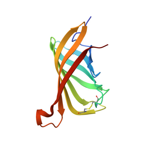
3T2X

Structure of shwanavidin low affinity mutant (F43A)


Experimental Data Snapshot

  • Method: X-RAY DIFFRACTION
  • Resolution: 1.15 Å
  • R-Value Free: 0.171 
  • R-Value Work: 0.145 
  • R-Value Observed: 0.146 

wwPDB Validation   3D Report Full Report


This is version 1.2 of the entry. See complete history


Literature

Structural Adaptation of a Thermostable Biotin-binding Protein in a Psychrophilic Environment.

Meir, A.Bayer, E.A.Livnah, O.

(2012) J Biol Chem 287: 17951-17962

  • DOI: https://doi.org/10.1074/jbc.M112.357186
  • Primary Citation of Related Structures:  
    3SZH, 3SZI, 3SZJ, 3T2W, 3T2X

  • PubMed Abstract: 

    Shwanavidin is an avidin-like protein from the marine proteobactrium Shewanella denitrificans, which exhibits an innate dimeric structure while maintaining high affinity toward biotin. A unique residue (Phe-43) from the L3,4 loop and a distinctive disulfide bridge were shown to account for the high affinity toward biotin. Phe-43 emulates the function and position of the critical intermonomeric Trp that characterizes the tetrameric avidins but is lacking in shwanavidin. The 18 copies of the apo-monomer revealed distinctive snapshots of L3,4 and Phe-43, providing rare insight into loop flexibility, binding site accessibility, and psychrophilic adaptation. Nevertheless, as in all avidins, shwanavidin also displays high thermostability properties. The unique features of shwanavidin may provide a platform for the design of a long sought after monovalent form of avidin, which would be ideal for novel types of biotechnological application.


  • Organizational Affiliation

    Department of Biological Chemistry, the Hebrew University of Jerusalem, Givat Ram, Jerusalem, Israel.


Macromolecules
Find similar proteins by:  (by identity cutoff)  |  3D Structure
Entity ID: 1
MoleculeChains Sequence LengthOrganismDetailsImage
Avidin/streptavidin
A, B, C, D
122Shewanella denitrificans OS217Mutation(s): 1 
Gene Names: Sden_0912
UniProt
Find proteins for Q12QS6 (Shewanella denitrificans (strain OS217 / ATCC BAA-1090 / DSM 15013))
Go to UniProtKB:  Q12QS6
Sequence Annotations
Expand
  • Reference Sequence
Experimental Data & Validation

Experimental Data

  • Method: X-RAY DIFFRACTION
  • Resolution: 1.15 Å
  • R-Value Free: 0.171 
  • R-Value Work: 0.145 
  • R-Value Observed: 0.146 
  • Space Group: H 3
Unit Cell:
Length ( Å )Angle ( ˚ )
a = 113.933α = 90
b = 113.933β = 90
c = 83.431γ = 120
Software Package:
Software NamePurpose
HKL-2000data collection
PHASERphasing
REFMACrefinement
HKL-2000data reduction
HKL-2000data scaling

Structure Validation

View Full Validation Report


Entry History 

Deposition Data

Revision History  (Full details and data files)

  • Version 1.0: 2012-04-11
    Type: Initial release
  • Version 1.1: 2012-04-25
    Changes: Database references
  • Version 1.2: 2012-06-13
    Changes: Database references