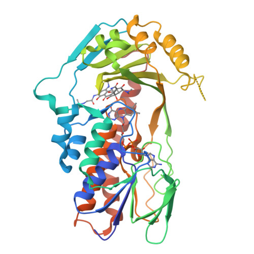
Crystal structure of TetX2 T280A: an adaptive mutant in complex with tigecycline
Walkiewicz, K., Shamoo, Y.To be published.
Experimental Data Snapshot
Starting Model: experimental
View more details
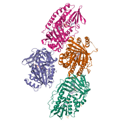
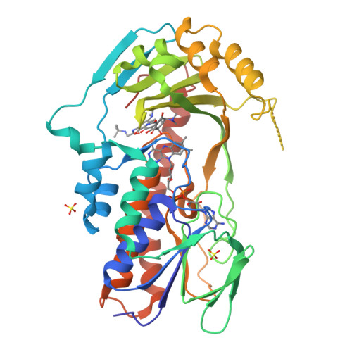
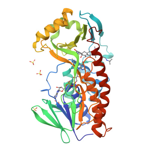
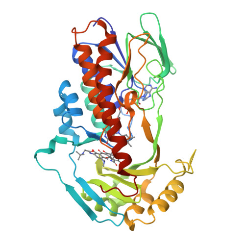
Entity ID: 1 | |||||
|---|---|---|---|---|---|
| Molecule | Chains | Sequence Length | Organism | Details | Image |
| TetX2 protein | 378 | Bacteroides thetaiotaomicron | Mutation(s): 3 Gene Names: tetX2 EC: 1.14.13.231 | 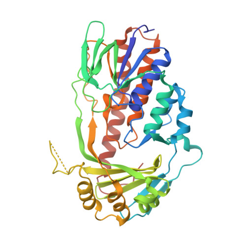 | |
UniProt | |||||
Find proteins for Q93L51 (Bacteroides thetaiotaomicron) Explore Q93L51 Go to UniProtKB: Q93L51 | |||||
Entity Groups | |||||
| Sequence Clusters | 30% Identity50% Identity70% Identity90% Identity95% Identity100% Identity | ||||
| UniProt Group | Q93L51 | ||||
Sequence AnnotationsExpand | |||||
| |||||
| Ligands 3 Unique | |||||
|---|---|---|---|---|---|
| ID | Chains | Name / Formula / InChI Key | 2D Diagram | 3D Interactions | |
| FAD Query on FAD | G [auth A], K [auth B], P [auth C], R [auth D] | FLAVIN-ADENINE DINUCLEOTIDE C27 H33 N9 O15 P2 VWWQXMAJTJZDQX-UYBVJOGSSA-N |  | ||
| T1C Query on T1C | H [auth A], M [auth B], O [auth C], Q [auth D] | TIGECYCLINE C29 H41 N5 O8 FPZLLRFZJZRHSY-HJYUBDRYSA-P |  | ||
| SO4 Query on SO4 | E [auth A] F [auth A] I [auth B] J [auth B] L [auth B] | SULFATE ION O4 S QAOWNCQODCNURD-UHFFFAOYSA-L |  | ||
| Length ( Å ) | Angle ( ˚ ) |
|---|---|
| a = 68.126 | α = 111.02 |
| b = 80.08 | β = 89.97 |
| c = 87.238 | γ = 92.97 |
| Software Name | Purpose |
|---|---|
| DENZO | data reduction |
| SCALEPACK | data scaling |
| PHENIX | refinement |
| PDB_EXTRACT | data extraction |
| JBluIce-EPICS | data collection |
| HKL-2000 | data reduction |
| HKL-2000 | data scaling |
| PHASER | phasing |
RCSB PDB is hosted by

RCSB PDB is a member of the