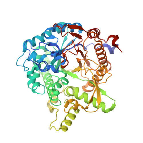
3VIF

Crystal structure of beta-glucosidase from termite Neotermes koshunensis in complex with gluconolactone


Experimental Data Snapshot

  • Method: X-RAY DIFFRACTION
  • Resolution: 1.00 Å
  • R-Value Free: 0.142 
  • R-Value Work: 0.122 
  • R-Value Observed: 0.123 

Starting Model: experimental
View more details

wwPDB Validation   3D Report Full Report


Ligand Structure Quality Assessment 


This is version 1.3 of the entry. See complete history


Literature

High-resolution structures of Neotermes koshunensis beta-glucosidase mutants provide insights into the catalytic mechanism and the synthesis of glucoconjugates

Jeng, W.Y.Wang, N.C.Lin, C.T.Chang, W.J.Liu, C.I.Wang, A.H.J.

(2012) Acta Crystallogr D Biol Crystallogr 68: 829-838

  • DOI: https://doi.org/10.1107/S0907444912013224
  • Primary Citation of Related Structures:  
    3VIF, 3VIG, 3VIH, 3VII, 3VIJ, 3VIK, 3VIL, 3VIM, 3VIN, 3VIO, 3VIP

  • PubMed Abstract: 

    NkBgl, a β-glucosidase from Neotermes koshunensis, is a β-retaining glycosyl hydrolase family 1 enzyme that cleaves β-glucosidic linkages in disaccharide or glucose-substituted molecules. β-Glucosidases have been widely used in several applications. For example, mutagenesis of the attacking nucleophile in β-glucosidase has been conducted to convert it into a glycosynthase for the synthesis of oligosaccharides. Here, several high-resolution structures of wild-type or mutated NkBgl in complex with different ligand molecules are reported. In the wild-type NkBgl structures it was found that glucose-like glucosidase inhibitors bind to the glycone-binding pocket, allowing the buffer molecule HEPES to remain in the aglycone-binding pocket. In the crystal structures of NkBgl E193A, E193S and E193D mutants Glu193 not only acts as the catalytic acid/base but also plays an important role in controlling substrate entry and product release. Furthermore, in crystal structures of the NkBgl E193D mutant it was found that new glucoconjugates were generated by the conjugation of glucose (hydrolyzed product) and HEPES/EPPS/opipramol (buffer components). Based on the wild-type and E193D-mutant structures of NkBgl, the glucosidic bond of cellobiose or salicin was hydrolyzed and a new bond was subsequently formed between glucose and HEPES/EPPS/opipramol to generate new glucopyranosidic products through the transglycosylation reaction in the NkBgl E193D mutant. This finding highlights an innovative way to further improve β-glucosidases for the enzymatic synthesis of oligosaccharides.


  • Organizational Affiliation

    Institute of Biological Chemistry, Academia Sinica, Taipei 115, Taiwan.


Macromolecules
Find similar proteins by:  (by identity cutoff)  |  3D Structure
Entity ID: 1
MoleculeChains Sequence LengthOrganismDetailsImage
Beta-glucosidase487Neotermes koshunensisMutation(s): 0 
Gene Names: NkBG
EC: 3.2.1.21
UniProt
Find proteins for Q8T0W7 (Neotermes koshunensis)
Explore Q8T0W7 
Go to UniProtKB:  Q8T0W7
Entity Groups  
Sequence Clusters30% Identity50% Identity70% Identity90% Identity95% Identity100% Identity
UniProt GroupQ8T0W7
Sequence Annotations
Expand
  • Reference Sequence
Small Molecules
Ligands 5 Unique
IDChains Name / Formula / InChI Key2D Diagram3D Interactions
EPE
Query on EPE

Download Ideal Coordinates CCD File 
C [auth A]4-(2-HYDROXYETHYL)-1-PIPERAZINE ETHANESULFONIC ACID
C8 H18 N2 O4 S
JKMHFZQWWAIEOD-UHFFFAOYSA-N
LGC
Query on LGC

Download Ideal Coordinates CCD File 
B [auth A]D-glucono-1,5-lactone
C6 H10 O6
PHOQVHQSTUBQQK-SQOUGZDYSA-N
GOL
Query on GOL

Download Ideal Coordinates CCD File 
D [auth A]GLYCEROL
C3 H8 O3
PEDCQBHIVMGVHV-UHFFFAOYSA-N
CL
Query on CL

Download Ideal Coordinates CCD File 
F [auth A],
G [auth A]
CHLORIDE ION
Cl
VEXZGXHMUGYJMC-UHFFFAOYSA-M
NA
Query on NA

Download Ideal Coordinates CCD File 
E [auth A]SODIUM ION
Na
FKNQFGJONOIPTF-UHFFFAOYSA-N
Experimental Data & Validation

Experimental Data

  • Method: X-RAY DIFFRACTION
  • Resolution: 1.00 Å
  • R-Value Free: 0.142 
  • R-Value Work: 0.122 
  • R-Value Observed: 0.123 
  • Space Group: C 1 2 1
Unit Cell:
Length ( Å )Angle ( ˚ )
a = 92.95α = 90
b = 68.479β = 95.33
c = 74.846γ = 90
Software Package:
Software NamePurpose
HKL-2000data collection
MOLREPphasing
REFMACrefinement
HKL-2000data reduction
HKL-2000data scaling

Structure Validation

View Full Validation Report


Ligand Structure Quality Assessment 


Entry History 

Deposition Data

Revision History  (Full details and data files)

  • Version 1.0: 2012-07-04
    Type: Initial release
  • Version 1.1: 2013-06-26
    Changes: Database references
  • Version 1.2: 2020-07-29
    Type: Remediation
    Reason: Carbohydrate remediation
    Changes: Database references, Derived calculations, Structure summary
  • Version 1.3: 2023-11-08
    Changes: Data collection, Database references, Refinement description, Structure summary