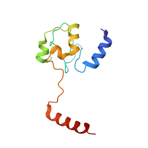
3WUI

Dimeric horse cytochrome c formed by refolding from molten globule state


Experimental Data Snapshot

  • Method: X-RAY DIFFRACTION
  • Resolution: 1.80 Å
  • R-Value Free: 0.227 
  • R-Value Work: 0.181 
  • R-Value Observed: 0.183 

Starting Model: experimental
View more details

wwPDB Validation   3D Report Full Report


Ligand Structure Quality Assessment 


This is version 2.2 of the entry. See complete history


Literature

Formation of domain-swapped oligomer of cytochrome C from its molten globule state oligomer.

Deshpande, M.S.Parui, P.P.Kamikubo, H.Yamanaka, M.Nagao, S.Komori, H.Kataoka, M.Higuchi, Y.Hirota, S.

(2014) Biochemistry 53: 4696-4703

  • DOI: https://doi.org/10.1021/bi500497s
  • Primary Citation of Related Structures:  
    3WUI

  • PubMed Abstract: 

    Many proteins, including cytochrome c (cyt c), have been shown to form domain-swapped oligomers, but the factors governing the oligomerization process remain unrevealed. We obtained oligomers of cyt c by refolding cyt c from its acid molten globule state to neutral pH state under high protein and ion concentrations. The amount of oligomeric cyt c obtained depended on the nature of the anion (chaotropic or kosmotropic) in the solution: ClO4(-) (oligomers, 11% ± 2% (heme unit)), SCN(-) (10% ± 2%), I(-) (6% ± 2%), NO3(-) (3% ± 1%), Br(-) (2% ± 1%), Cl(-) (2% ± 1%), and SO4(2-) (3% ± 1%) for refolding of 2 mM cyt c (anion concentration 125 mM). Dimeric cyt c obtained by refolding from the molten globule state exhibited a domain-swapped structure, in which the C-terminal α-helices were exchanged between protomers. According to small-angle X-ray scattering measurements, approximately 25% of the cyt c molecules were dimerized in the molten globule state containing 125 mM ClO4(-). These results indicate that a certain amount of molten globule state oligomers of cyt c convert to domain-swapped oligomers during refolding and that the intermolecular interactions necessary for domain swapping are present in the molten globule state.


  • Organizational Affiliation

    Graduate School of Materials Science, Nara Institute of Science and Technology , 8916-5 Takayama, Ikoma, Nara 630-0192, Japan.


Macromolecules
Find similar proteins by:  (by identity cutoff)  |  3D Structure
Entity ID: 1
MoleculeChains Sequence LengthOrganismDetailsImage
Cytochrome c104Equus caballusMutation(s): 0 
UniProt
Find proteins for P00004 (Equus caballus)
Explore P00004 
Go to UniProtKB:  P00004
Entity Groups  
Sequence Clusters30% Identity50% Identity70% Identity90% Identity95% Identity100% Identity
UniProt GroupP00004
Sequence Annotations
Expand
  • Reference Sequence
Small Molecules
Experimental Data & Validation

Experimental Data

  • Method: X-RAY DIFFRACTION
  • Resolution: 1.80 Å
  • R-Value Free: 0.227 
  • R-Value Work: 0.181 
  • R-Value Observed: 0.183 
  • Space Group: C 2 2 21
Unit Cell:
Length ( Å )Angle ( ˚ )
a = 60.948α = 90
b = 94.145β = 90
c = 36.39γ = 90
Software Package:
Software NamePurpose
HKL-2000data collection
REFMACrefinement
HKL-2000data reduction
HKL-2000data scaling
REFMACphasing

Structure Validation

View Full Validation Report


Ligand Structure Quality Assessment 


Entry History 

Deposition Data

Revision History  (Full details and data files)

  • Version 1.0: 2014-07-16
    Type: Initial release
  • Version 2.0: 2019-10-02
    Changes: Atomic model, Data collection, Derived calculations, Non-polymer description, Structure summary
  • Version 2.1: 2022-08-24
    Changes: Database references, Derived calculations
  • Version 2.2: 2023-11-08
    Changes: Data collection, Refinement description