3ZRQ

Crystal structure and substrate specificity of a thermophilic archaeal serine : pyruvate aminotransferase from Sulfolobus solfataricus


Experimental Data Snapshot

  • Method: X-RAY DIFFRACTION
  • Resolution: 1.80 Å
  • R-Value Free: 0.234 
  • R-Value Work: 0.189 
  • R-Value Observed: 0.191 

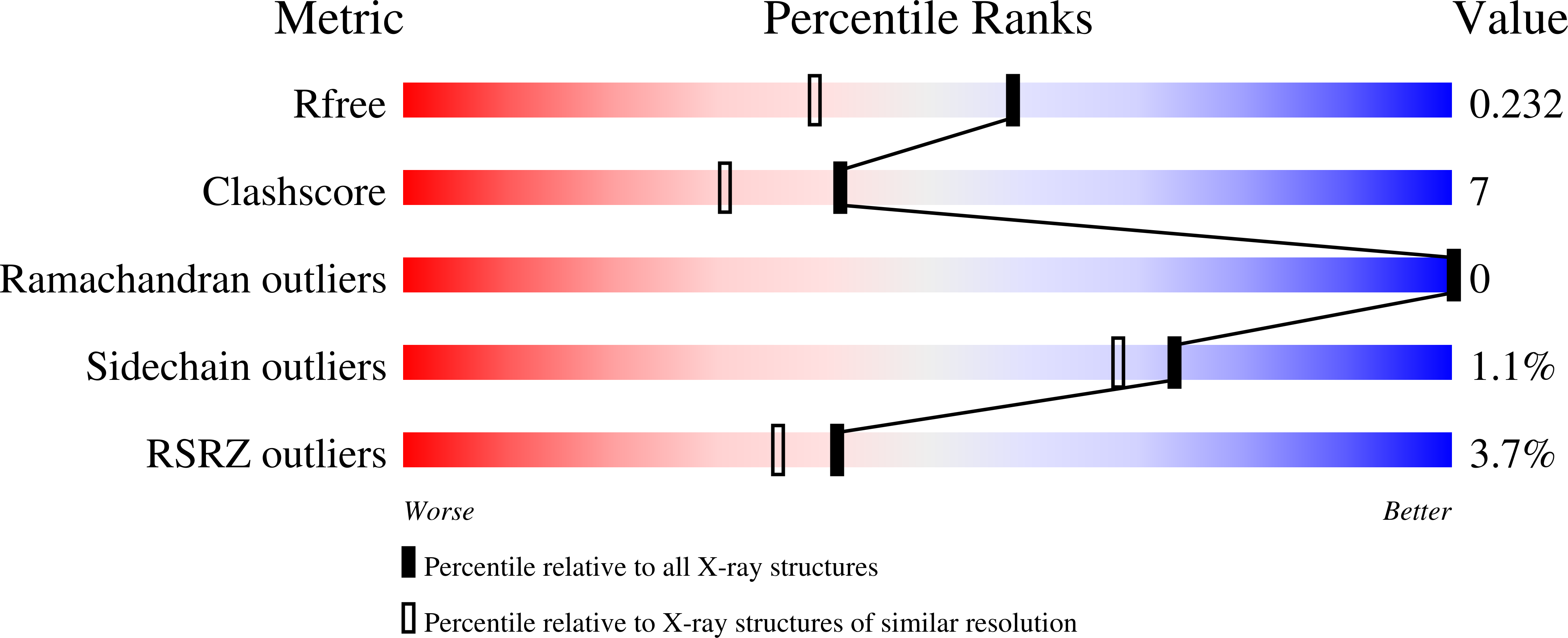
wwPDB Validation   3D Report Full Report


Ligand Structure Quality Assessment 


This is version 1.2 of the entry. See complete history


Literature

Crystal Structure and Substrate Specificity of the Thermophilic Serine:Pyruvate Aminotransferase from Sulfolobus Solfataricus

Sayer, C.Bommer, M.Isupov, M.N.Ward, J.Littlechild, J.

(2012) Acta Crystallogr D Biol Crystallogr 68: 763

  • DOI: https://doi.org/10.1107/S0907444912011274
  • Primary Citation of Related Structures:  
    3ZRP, 3ZRQ, 3ZRR

  • PubMed Abstract: 

    The three-dimensional structure of the Sulfolobus solfataricus serine:pyruvate aminotransferase has been determined to 1.8 Å resolution. The structure of the protein is a homodimer that adopts the type I fold of pyridoxal 5'-phosphate (PLP)-dependent aminotransferases. The structure revealed the PLP cofactor covalently bound in the active site to the active-site lysine in the internal aldimine form. The structure of the S. solfataricus enzyme was also determined with an amino form of the cofactor pyridoxamine 5'-phosphate bound in the active site and in complex with gabaculine, an aminotransferase inhibitor. These structures showed the changes in the enzyme active site during the course of the catalytic reaction. A comparison of the structure of the S. solfataricus enzyme with that of the closely related alanine:glyoxylate aminotransferase has identified structural features that are proposed to be responsible for the differences in substrate specificity between the two enzymes. These results have been complemented by biochemical studies of the substrate specificity and thermostability of the S. solfataricus enzyme.


  • Organizational Affiliation

    Henry Wellcome Building for Biocatalysis, Biosciences, College of Life and Environmental Sciences, University of Exeter, Stocker Road, Exeter EX4 4QD, England.


Macromolecules
Find similar proteins by:  (by identity cutoff)  |  3D Structure
Entity ID: 1
MoleculeChains Sequence LengthOrganismDetailsImage
SERINE-PYRUVATE AMINOTRANSFERASE (AGXT)
A, B
384Saccharolobus solfataricus P2Mutation(s): 0 
EC: 2.6.1.51
UniProt
Find proteins for Q97VM5 (Saccharolobus solfataricus (strain ATCC 35092 / DSM 1617 / JCM 11322 / P2))
Explore Q97VM5 
Go to UniProtKB:  Q97VM5
Entity Groups  
Sequence Clusters30% Identity50% Identity70% Identity90% Identity95% Identity100% Identity
UniProt GroupQ97VM5
Sequence Annotations
Expand
  • Reference Sequence
Experimental Data & Validation

Experimental Data

  • Method: X-RAY DIFFRACTION
  • Resolution: 1.80 Å
  • R-Value Free: 0.234 
  • R-Value Work: 0.189 
  • R-Value Observed: 0.191 
  • Space Group: C 1 2 1
Unit Cell:
Length ( Å )Angle ( ˚ )
a = 160.13α = 90
b = 55.05β = 111.03
c = 101.59γ = 90
Software Package:
Software NamePurpose
REFMACrefinement
MOSFLMdata reduction
SCALAdata scaling
MOLREPphasing

Structure Validation

View Full Validation Report


Ligand Structure Quality Assessment 


Entry History 

Deposition Data

Revision History  (Full details and data files)

  • Version 1.0: 2012-06-27
    Type: Initial release
  • Version 1.1: 2012-07-11
    Changes: Other
  • Version 1.2: 2024-05-08
    Changes: Data collection, Database references, Derived calculations, Other