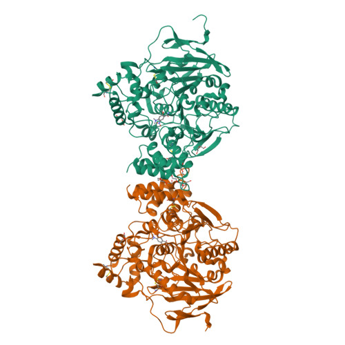
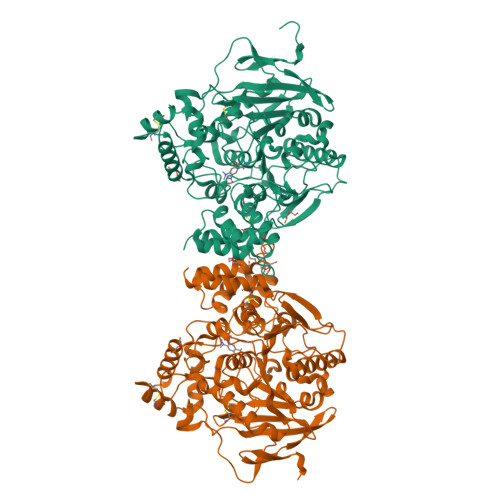
4A23

Mus musculus Acetylcholinesterase in complex with racemic C5685


Experimental Data Snapshot

  • Method: X-RAY DIFFRACTION
  • Resolution: 2.40 Å
  • R-Value Free: 0.209 
  • R-Value Work: 0.174 
  • R-Value Observed: 0.175 

Starting Model: experimental
View more details

wwPDB Validation   3D Report Full Report


Ligand Structure Quality Assessment 


This is version 1.4 of the entry. See complete history


Literature

Targeting Acetylcholinesterase: Identification of Chemical Leads by High Throughput Screening, Structure Determination and Molecular Modeling.

Berg, L.Andersson, C.D.Artursson, E.Hornberg, A.Tunemalm, A.K.Linusson, A.Ekstrom, F.

(2011) PLoS One 6: 26039

  • DOI: https://doi.org/10.1371/journal.pone.0026039
  • Primary Citation of Related Structures:  
    4A23

  • PubMed Abstract: 

    Acetylcholinesterase (AChE) is an essential enzyme that terminates cholinergic transmission by rapid hydrolysis of the neurotransmitter acetylcholine. Compounds inhibiting this enzyme can be used (inter alia) to treat cholinergic deficiencies (e.g. in Alzheimer's disease), but may also act as dangerous toxins (e.g. nerve agents such as sarin). Treatment of nerve agent poisoning involves use of antidotes, small molecules capable of reactivating AChE. We have screened a collection of organic molecules to assess their ability to inhibit the enzymatic activity of AChE, aiming to find lead compounds for further optimization leading to drugs with increased efficacy and/or decreased side effects. 124 inhibitors were discovered, with considerable chemical diversity regarding size, polarity, flexibility and charge distribution. An extensive structure determination campaign resulted in a set of crystal structures of protein-ligand complexes. Overall, the ligands have substantial interactions with the peripheral anionic site of AChE, and the majority form additional interactions with the catalytic site (CAS). Reproduction of the bioactive conformation of six of the ligands using molecular docking simulations required modification of the default parameter settings of the docking software. The results show that docking-assisted structure-based design of AChE inhibitors is challenging and requires crystallographic support to obtain reliable results, at least with currently available software. The complex formed between C5685 and Mus musculus AChE (C5685•mAChE) is a representative structure for the general binding mode of the determined structures. The CAS binding part of C5685 could not be structurally determined due to a disordered electron density map and the developed docking protocol was used to predict the binding modes of this part of the molecule. We believe that chemical modifications of our discovered inhibitors, biochemical and biophysical characterization, crystallography and computational chemistry provide a route to novel AChE inhibitors and reactivators.


  • Organizational Affiliation

    Department of Chemistry, Umeå University, Umeå, Sweden.


Macromolecules
Find similar proteins by:  (by identity cutoff)  |  3D Structure
Entity ID: 1
MoleculeChains Sequence LengthOrganismDetailsImage
ACETYLCHOLINESTERASE
A, B
548Mus musculusMutation(s): 0 
EC: 3.1.1.7
UniProt & NIH Common Fund Data Resources
Find proteins for P21836 (Mus musculus)
Explore P21836 
Go to UniProtKB:  P21836
IMPC:  MGI:87876
Entity Groups  
Sequence Clusters30% Identity50% Identity70% Identity90% Identity95% Identity100% Identity
UniProt GroupP21836
Sequence Annotations
Expand
  • Reference Sequence
Small Molecules
Ligands 4 Unique
IDChains Name / Formula / InChI Key2D Diagram3D Interactions
PE4
Query on PE4

Download Ideal Coordinates CCD File 
K [auth B]2-{2-[2-(2-{2-[2-(2-ETHOXY-ETHOXY)-ETHOXY]-ETHOXY}-ETHOXY)-ETHOXY]-ETHOXY}-ETHANOL
C16 H34 O8
PJWQOENWHPEPKI-UHFFFAOYSA-N
C57
Query on C57

Download Ideal Coordinates CCD File 
D [auth A],
I [auth B]
4-(DIMETHYLAMINO)-N-{[(2S)-1-ETHYLPYRROLIDIN-2-YL]METHYL}-2-METHOXY-5-NITROBENZAMIDE
C17 H26 N4 O4
AFNPLUMDSLEAPD-LBPRGKRZSA-N
C56
Query on C56

Download Ideal Coordinates CCD File 
C [auth A],
H [auth B]
4-(DIMETHYLAMINO)-N-{[(2R)-1-ETHYLPYRROLIDIN-2-YL]METHYL}-2-METHOXY-5-NITROBENZAMIDE
C17 H26 N4 O4
AFNPLUMDSLEAPD-GFCCVEGCSA-N
PEG
Query on PEG

Download Ideal Coordinates CCD File 
E [auth A],
F [auth A],
G [auth A],
J [auth B],
L [auth B]
DI(HYDROXYETHYL)ETHER
C4 H10 O3
MTHSVFCYNBDYFN-UHFFFAOYSA-N
Binding Affinity Annotations 
IDSourceBinding Affinity
C56 PDBBind:  4A23 IC50: 1300 (nM) from 1 assay(s)
Experimental Data & Validation

Experimental Data

  • Method: X-RAY DIFFRACTION
  • Resolution: 2.40 Å
  • R-Value Free: 0.209 
  • R-Value Work: 0.174 
  • R-Value Observed: 0.175 
  • Space Group: P 21 21 21
Unit Cell:
Length ( Å )Angle ( ˚ )
a = 78.923α = 90
b = 111.828β = 90
c = 226.999γ = 90
Software Package:
Software NamePurpose
PHENIXrefinement
XDSdata reduction
SCALAdata scaling
REFMACphasing

Structure Validation

View Full Validation Report


Ligand Structure Quality Assessment 


Entry History 

Deposition Data

Revision History  (Full details and data files)

  • Version 1.0: 2011-12-07
    Type: Initial release
  • Version 1.1: 2011-12-28
    Changes: Other
  • Version 1.2: 2018-01-17
    Changes: Data collection
  • Version 1.3: 2019-10-16
    Changes: Data collection, Other
  • Version 1.4: 2023-12-20
    Changes: Data collection, Database references, Derived calculations, Refinement description