4AAY | pdb_00004aay

Crystal Structure of the arsenite oxidase protein complex from Rhizobium species strain NT-26


Experimental Data Snapshot

  • Method: X-RAY DIFFRACTION
  • Resolution: 2.70 Å
  • R-Value Free: 
    0.211 (Depositor), 0.210 (DCC) 
  • R-Value Work: 
    0.193 (Depositor), 0.190 (DCC) 
  • R-Value Observed: 
    0.194 (Depositor) 

Starting Model: experimental
View more details

wwPDB Validation   3D Report Full Report


Ligand Structure Quality Assessment 

Created with Raphaël 2.3.0Worse 01 BetterLigand structure goodness of fit to experimental dataBest fitted MGDClick on this verticalbar to view details

This is version 1.4 of the entry. See complete history


Literature

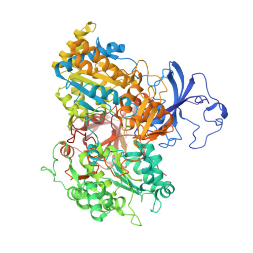
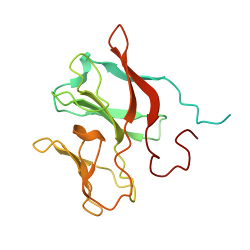
The Respiratory Arsenite Oxidase: Structure and the Role of Residues Surrounding the Rieske Cluster.

Warelow, T.P.Oke, M.Schoepp-Cothenet, B.Dahl, J.U.Bruselat, N.Sivalingam, G.N.Leimkuhler, S.Thalassinos, K.Kappler, U.Naismith, J.H.Santini, J.M.

(2013) PLoS One 8: 72535

  • DOI: https://doi.org/10.1371/journal.pone.0072535
  • Primary Citation of Related Structures:  
    4AAY

  • PubMed Abstract: 

    The arsenite oxidase (Aio) from the facultative autotrophic Alphaproteobacterium Rhizobium sp. NT-26 is a bioenergetic enzyme involved in the oxidation of arsenite to arsenate. The enzyme from the distantly related heterotroph, Alcaligenes faecalis, which is thought to oxidise arsenite for detoxification, consists of a large α subunit (AioA) with bis-molybdopterin guanine dinucleotide at its active site and a 3Fe-4S cluster, and a small β subunit (AioB) which contains a Rieske 2Fe-2S cluster. The successful heterologous expression of the NT-26 Aio in Escherichia coli has resulted in the solution of its crystal structure. The NT-26 Aio, a heterotetramer, shares high overall similarity to the heterodimeric arsenite oxidase from A. faecalis but there are striking differences in the structure surrounding the Rieske 2Fe-2S cluster which we demonstrate explains the difference in the observed redox potentials (+225 mV vs. +130/160 mV, respectively). A combination of site-directed mutagenesis and electron paramagnetic resonance was used to explore the differences observed in the structure and redox properties of the Rieske cluster. In the NT-26 AioB the substitution of a serine (S126 in NT-26) for a threonine as in the A. faecalis AioB explains a -20 mV decrease in redox potential. The disulphide bridge in the A. faecalis AioB which is conserved in other betaproteobacterial AioB subunits and the Rieske subunit of the cytochrome bc 1 complex is absent in the NT-26 AioB subunit. The introduction of a disulphide bridge had no effect on Aio activity or protein stability but resulted in a decrease in the redox potential of the cluster. These results are in conflict with previous data on the betaproteobacterial AioB subunit and the Rieske of the bc 1 complex where removal of the disulphide bridge had no effect on the redox potential of the former but a decrease in cluster stability was observed in the latter.


  • Organizational Affiliation

    Institute of Structural and Molecular Biology, University College London, London, United Kingdom.


Macromolecules
Find similar proteins by:  (by identity cutoff)  |  3D Structure
Entity ID: 1
MoleculeChains Sequence LengthOrganismDetailsImage
AROA
A, C, E, G
845Pseudorhizobium banfieldiaeMutation(s): 0 
EC: 1.20.98.1
UniProt
Find proteins for Q6VAL8 (Pseudorhizobium banfieldiae)
Explore Q6VAL8 
Go to UniProtKB:  Q6VAL8
Entity Groups  
Sequence Clusters30% Identity50% Identity70% Identity90% Identity95% Identity100% Identity
UniProt GroupQ6VAL8
Sequence Annotations
Expand
  • Reference Sequence
Find similar proteins by:  (by identity cutoff)  |  3D Structure
Entity ID: 2
MoleculeChains Sequence LengthOrganismDetailsImage
AROB
B, D, F, H
175Pseudorhizobium banfieldiaeMutation(s): 0 
UniProt
Find proteins for Q6VAL9 (Pseudorhizobium banfieldiae)
Explore Q6VAL9 
Go to UniProtKB:  Q6VAL9
Entity Groups  
Sequence Clusters30% Identity50% Identity70% Identity90% Identity95% Identity100% Identity
UniProt GroupQ6VAL9
Sequence Annotations
Expand
  • Reference Sequence
Small Molecules
Ligands 5 Unique
IDChains Name / Formula / InChI Key2D Diagram3D Interactions
MGD
Query on MGD

Download Ideal Coordinates CCD File 
AA [auth G]
BA [auth G]
I [auth A]
J [auth A]
O [auth C]
2-AMINO-5,6-DIMERCAPTO-7-METHYL-3,7,8A,9-TETRAHYDRO-8-OXA-1,3,9,10-TETRAAZA-ANTHRACEN-4-ONE GUANOSINE DINUCLEOTIDE
C20 H26 N10 O13 P2 S2
VQAGYJCYOLHZDH-ILXWUORBSA-N
F3S
Query on F3S

Download Ideal Coordinates CCD File 
EA [auth G],
M [auth A],
S [auth C],
Y [auth E]
FE3-S4 CLUSTER
Fe3 S4
FCXHZBQOKRZXKS-UHFFFAOYSA-N
FES
Query on FES

Download Ideal Coordinates CCD File 
FA [auth H],
N [auth B],
T [auth D],
Z [auth F]
FE2/S2 (INORGANIC) CLUSTER
Fe2 S2
NIXDOXVAJZFRNF-UHFFFAOYSA-N
4MO
Query on 4MO

Download Ideal Coordinates CCD File 
DA [auth G],
L [auth A],
R [auth C],
X [auth E]
MOLYBDENUM(IV) ION
Mo
ZIKKVZAYJJZBGE-UHFFFAOYSA-N
O
Query on O

Download Ideal Coordinates CCD File 
CA [auth G],
K [auth A],
Q [auth C],
W [auth E]
OXYGEN ATOM
O
XLYOFNOQVPJJNP-UHFFFAOYSA-N
Experimental Data & Validation

Experimental Data

  • Method: X-RAY DIFFRACTION
  • Resolution: 2.70 Å
  • R-Value Free:  0.211 (Depositor), 0.210 (DCC) 
  • R-Value Work:  0.193 (Depositor), 0.190 (DCC) 
  • R-Value Observed: 0.194 (Depositor) 
Space Group: P 21 21 2
Unit Cell:
Length ( Å )Angle ( ˚ )
a = 149.13α = 90
b = 232.96β = 90
c = 141.87γ = 90
Software Package:
Software NamePurpose
REFMACrefinement
xia2data reduction
xia2data scaling
PHASERphasing

Structure Validation

View Full Validation Report


Ligand Structure Quality Assessment 

Created with Raphaël 2.3.0Worse 01 BetterLigand structure goodness of fit to experimental dataBest fitted MGDClick on this verticalbar to view details

Entry History 

Deposition Data

Revision History  (Full details and data files)

  • Version 1.0: 2012-12-12
    Type: Initial release
  • Version 1.1: 2013-09-25
    Changes: Database references
  • Version 1.2: 2013-11-20
    Changes: Derived calculations, Refinement description
  • Version 1.3: 2014-03-12
    Changes: Source and taxonomy
  • Version 1.4: 2023-12-20
    Changes: Data collection, Database references, Derived calculations, Other, Refinement description