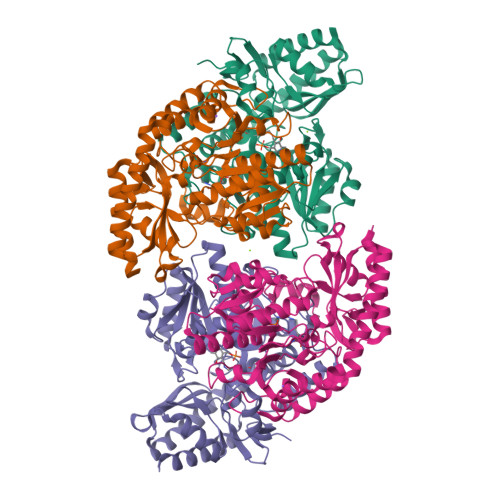
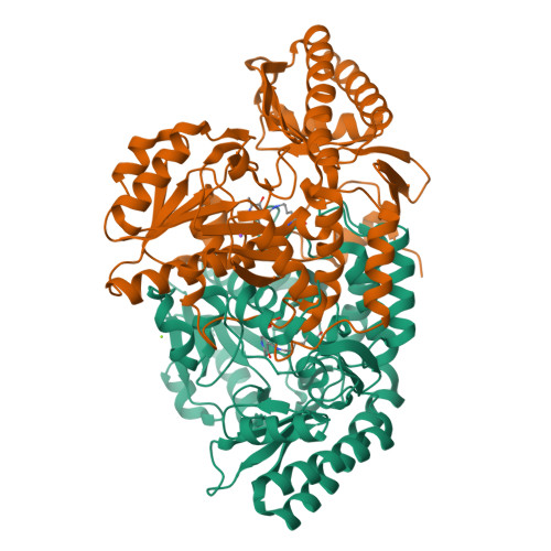
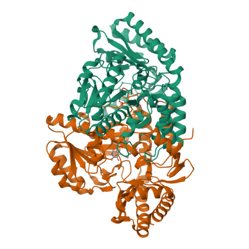
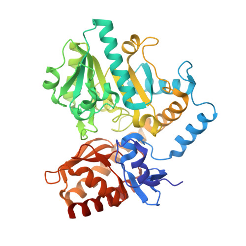
4ADB

Structural and functional study of succinyl-ornithine transaminase from E. coli


Experimental Data Snapshot

  • Method: X-RAY DIFFRACTION
  • Resolution: 2.20 Å
  • R-Value Free: 0.219 
  • R-Value Work: 0.171 
  • R-Value Observed: 0.173 

Starting Model: experimental
View more details

wwPDB Validation   3D Report Full Report


Ligand Structure Quality Assessment 


This is version 1.2 of the entry. See complete history


Literature

Determination of the Structure of the Catabolic N-Succinylornithine Transaminase (Astc) from Escherichia Coli.

Newman, J.Seabrook, S.Surjadi, R.Williams, C.C.Lucent, D.Wilding, M.Scott, C.Peat, T.S.

(2013) PLoS One 8: 58298

  • DOI: https://doi.org/10.1371/journal.pone.0058298
  • Primary Citation of Related Structures:  
    4ADB, 4ADC, 4ADD, 4ADE

  • PubMed Abstract: 

    Escherichia coli possesses two acyl ornithine aminotransferases, one catabolic (AstC) and the other anabolic (ArgD), that participate in L-arginine metabolism. Although only 58% identical, the enzymes have been shown to be functionally interchangeable. Here we have purified AstC and have obtained X-ray crystal structures of apo and holo-AstC and of the enzyme complexed with its physiological substrate, succinylornithine. We compare the structures obtained in this study with those of ArgD from Salmonella typhimurium obtained elsewhere, finding several notable differences. Docking studies were used to explore the docking modes of several substrates (ornithine, succinylornithine and acetylornithine) and the co-substrate glutamate/α-ketogluterate. The docking studies support our observations that AstC has a strong preference for acylated ornithine species over ornithine itself, and suggest that the increase in specificity associated with acylation is caused by steric and desolvation effects rather than specific interactions between the substrate and enzyme.


  • Organizational Affiliation

    CSIRO Materials, Science and Engineering, Parkville, Australia.


Macromolecules
Find similar proteins by:  (by identity cutoff)  |  3D Structure
Entity ID: 1
MoleculeChains Sequence LengthOrganismDetailsImage
SUCCINYLORNITHINE TRANSAMINASE
A, B, C, D
406Escherichia coli BL21Mutation(s): 0 
EC: 2.6.1.17 (PDB Primary Data), 2.6.1.81 (PDB Primary Data)
UniProt
Find proteins for P77581 (Escherichia coli (strain K12))
Explore P77581 
Go to UniProtKB:  P77581
Entity Groups  
Sequence Clusters30% Identity50% Identity70% Identity90% Identity95% Identity100% Identity
UniProt GroupP77581
Sequence Annotations
Expand
  • Reference Sequence
Small Molecules
Ligands 3 Unique
IDChains Name / Formula / InChI Key2D Diagram3D Interactions
PLP
Query on PLP

Download Ideal Coordinates CCD File 
E [auth A],
H [auth B],
K [auth C],
M [auth D]
PYRIDOXAL-5'-PHOSPHATE
C8 H10 N O6 P
NGVDGCNFYWLIFO-UHFFFAOYSA-N
MG
Query on MG

Download Ideal Coordinates CCD File 
G [auth A],
J [auth B]
MAGNESIUM ION
Mg
JLVVSXFLKOJNIY-UHFFFAOYSA-N
NA
Query on NA

Download Ideal Coordinates CCD File 
F [auth A],
I [auth B],
L [auth C],
N [auth D]
SODIUM ION
Na
FKNQFGJONOIPTF-UHFFFAOYSA-N
Experimental Data & Validation

Experimental Data

  • Method: X-RAY DIFFRACTION
  • Resolution: 2.20 Å
  • R-Value Free: 0.219 
  • R-Value Work: 0.171 
  • R-Value Observed: 0.173 
  • Space Group: C 1 2 1
Unit Cell:
Length ( Å )Angle ( ˚ )
a = 184.708α = 90
b = 118.428β = 96.69
c = 109.792γ = 90
Software Package:
Software NamePurpose
REFMACrefinement
XDSdata reduction
SCALAdata scaling
PHASERphasing

Structure Validation

View Full Validation Report


Ligand Structure Quality Assessment 


Entry History 

Deposition Data

Revision History  (Full details and data files)

  • Version 1.0: 2013-01-16
    Type: Initial release
  • Version 1.1: 2013-03-27
    Changes: Database references
  • Version 1.2: 2023-12-20
    Changes: Data collection, Database references, Derived calculations, Other, Refinement description