4BEI | pdb_00004bei

V. cholera biofilm scaffolding protein RbmA in complex with 18-crown- 6


Experimental Data Snapshot

  • Method: X-RAY DIFFRACTION
  • Resolution: 2.60 Å
  • R-Value Free: 
    0.261 (Depositor), 0.260 (DCC) 
  • R-Value Work: 
    0.222 (Depositor), 0.220 (DCC) 
  • R-Value Observed: 
    0.224 (Depositor) 

Starting Model: experimental
View more details

wwPDB Validation   3D Report Full Report


Ligand Structure Quality Assessment 

Created with Raphaël 2.3.0Worse 01 BetterLigand structure goodness of fit to experimental dataBest fitted O4BClick on this verticalbar to view details

This is version 1.3 of the entry. See complete history


Literature

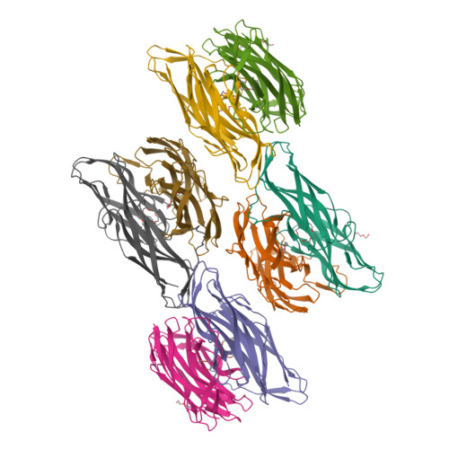
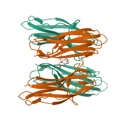
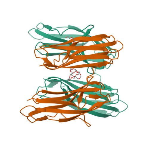
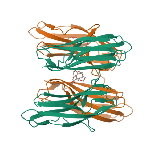
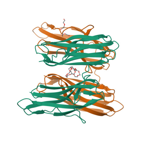
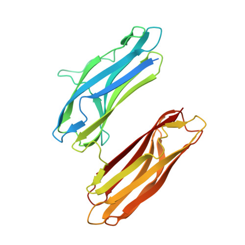
Structural Insights Into Rbma, a Biofilm Scaffolding Protein of V. Cholerae.

Maestre-Reyna, M.Wu, W.Wang, A.H.

(2013) PLoS One 8: 82458

  • DOI: https://doi.org/10.1371/journal.pone.0082458
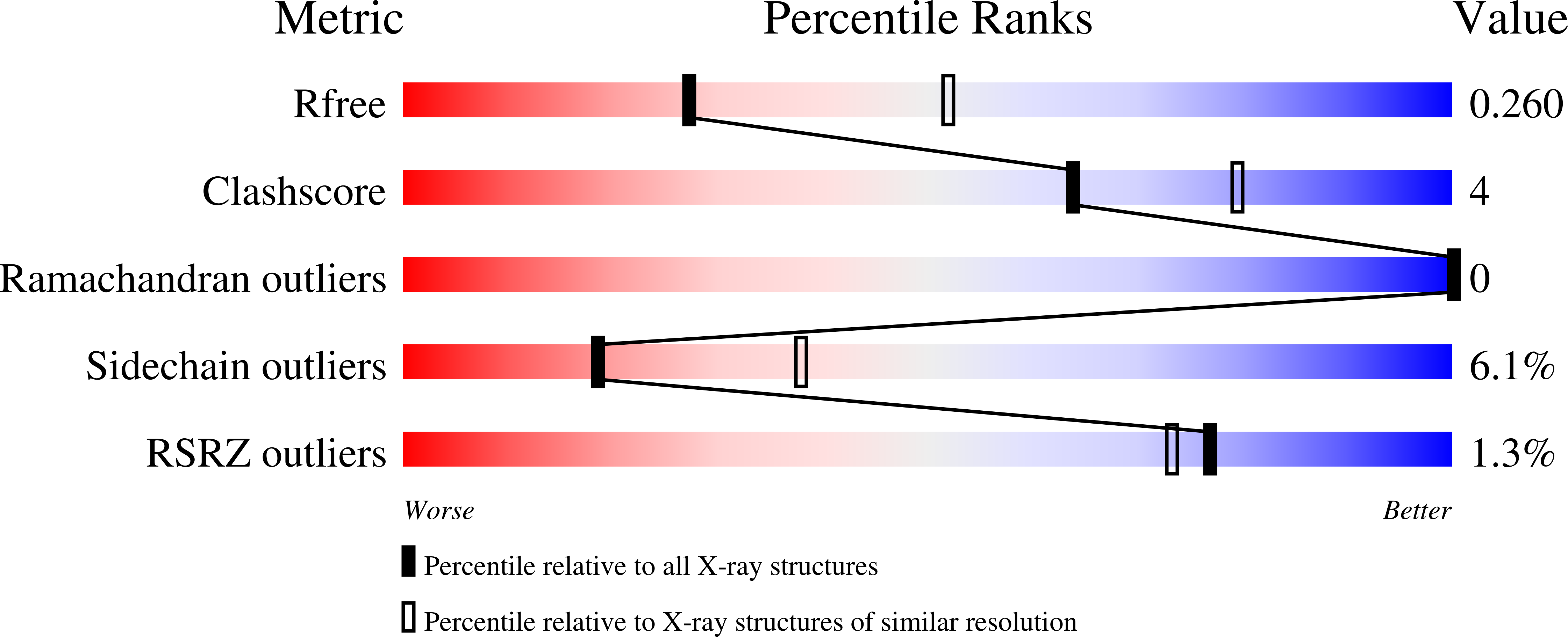
  • Primary Citation of Related Structures:  
    4BE5, 4BE6, 4BEI

  • PubMed Abstract: 

    V. cholerae can form sessile biofilms associated with abiotic surfaces, cyanobacteria, zoo-plankton, mollusks, or crustaceans. Along with the vibrio polysaccharide, secreted proteins of the rbm gene cluster are key to the biofilm ultrastructure. Here we provide a thorough structural characterization of RbmA, a protein involved in mediating cell-cell and cell-biofilm contacts. We correlate our structural findings with initial ligand specificity screening results, NMR protein-ligand interaction analysis, and complement our results with a full biocomputational study.


  • Organizational Affiliation

    Institute of Biological Chemistry, Academia Sinica, Taipei, Taiwan.


Macromolecules
Find similar proteins by:  (by identity cutoff)  |  3D Structure
Entity ID: 1
MoleculeChains Sequence LengthOrganismDetailsImage
RBMA
A, B, C, D, E
262Vibrio cholerae MJ-1236Mutation(s): 0 
Entity Groups  
Sequence Clusters30% Identity50% Identity70% Identity90% Identity95% Identity100% Identity
Sequence Annotations
Expand
  • Reference Sequence
Small Molecules
Ligands 2 Unique
IDChains Name / Formula / InChI Key2D Diagram3D Interactions
O4B
Query on O4B

Download Ideal Coordinates CCD File 
I [auth A]
K [auth B]
L [auth C]
M [auth D]
O [auth E]
1,4,7,10,13,16-HEXAOXACYCLOOCTADECANE
C12 H24 O6
XEZNGIUYQVAUSS-UHFFFAOYSA-N
PEG
Query on PEG

Download Ideal Coordinates CCD File 
J [auth A],
N [auth D],
P [auth E],
R [auth F]
DI(HYDROXYETHYL)ETHER
C4 H10 O3
MTHSVFCYNBDYFN-UHFFFAOYSA-N
Experimental Data & Validation

Experimental Data

  • Method: X-RAY DIFFRACTION
  • Resolution: 2.60 Å
  • R-Value Free:  0.261 (Depositor), 0.260 (DCC) 
  • R-Value Work:  0.222 (Depositor), 0.220 (DCC) 
  • R-Value Observed: 0.224 (Depositor) 
Space Group: P 31
Unit Cell:
Length ( Å )Angle ( ˚ )
a = 136.885α = 90
b = 136.885β = 90
c = 116.312γ = 120
Software Package:
Software NamePurpose
REFMACrefinement
HKL-2000data reduction
HKL-2000data scaling
PHASERphasing

Structure Validation

View Full Validation Report


Ligand Structure Quality Assessment 

Created with Raphaël 2.3.0Worse 01 BetterLigand structure goodness of fit to experimental dataBest fitted O4BClick on this verticalbar to view details

Entry History 

Deposition Data

Revision History  (Full details and data files)

  • Version 1.0: 2013-12-18
    Type: Initial release
  • Version 1.1: 2013-12-25
    Changes: Database references
  • Version 1.2: 2014-01-08
    Changes: Database references
  • Version 1.3: 2023-12-20
    Changes: Data collection, Database references, Derived calculations, Other, Refinement description