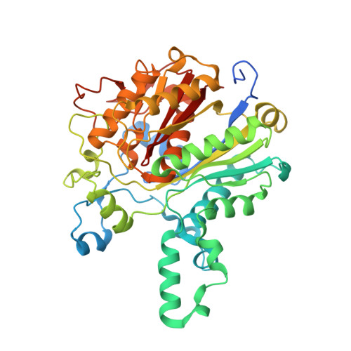
4C6U

Crystal structure of M. tuberculosis KasA in complex with TLM5


Experimental Data Snapshot

  • Method: X-RAY DIFFRACTION
  • Resolution: 2.40 Å
  • R-Value Free: 0.216 
  • R-Value Work: 0.164 
  • R-Value Observed: 0.167 

Starting Model: experimental
View more details

wwPDB Validation   3D Report Full Report


Ligand Structure Quality Assessment 


This is version 1.4 of the entry. See complete history


Literature

Structural Basis for the Recognition of Mycolic Acid Precursors by Kasa, a Condensing Enzyme and Drug Target from Mycobacterium Tuberculosis

Schiebel, J.Kapilashrami, K.Fekete, A.Bommineni, G.R.Schaefer, C.M.Mueller, M.J.Tonge, P.J.Kisker, C.

(2013) J Biol Chem 288: 34190

  • DOI: https://doi.org/10.1074/jbc.M113.511436
  • Primary Citation of Related Structures:  
    4C6U, 4C6V, 4C6W, 4C6X, 4C6Z, 4C70, 4C71, 4C72, 4C73

  • PubMed Abstract: 

    The survival of Mycobacterium tuberculosis depends on mycolic acids, very long α-alkyl-β-hydroxy fatty acids comprising 60-90 carbon atoms. However, despite considerable efforts, little is known about how enzymes involved in mycolic acid biosynthesis recognize and bind their hydrophobic fatty acyl substrates. The condensing enzyme KasA is pivotal for the synthesis of very long (C38-42) fatty acids, the precursors of mycolic acids. To probe the mechanism of substrate and inhibitor recognition by KasA, we determined the structure of this protein in complex with a mycobacterial phospholipid and with several thiolactomycin derivatives that were designed as substrate analogs. Our structures provide consecutive snapshots along the reaction coordinate for the enzyme-catalyzed reaction and support an induced fit mechanism in which a wide cavity is established through the concerted opening of three gatekeeping residues and several α-helices. The stepwise characterization of the binding process provides mechanistic insights into the induced fit recognition in this system and serves as an excellent foundation for the development of high affinity KasA inhibitors.


  • Organizational Affiliation

    Rudolf Virchow Center for Experimental Biomedicine, Institute for Structural Biology, University of Wuerzburg, D-97080 Wuerzburg, Germany; Institute of Pharmacy and Food Chemistry, University of Wuerzburg, Am Hubland, D-97074 Wuerzburg, Germany.


Macromolecules
Find similar proteins by:  (by identity cutoff)  |  3D Structure
Entity ID: 1
MoleculeChains Sequence LengthOrganismDetailsImage
3-OXOACYL-[ACYL-CARRIER-PROTEIN] SYNTHASE 1439Mycobacterium tuberculosis H37RvMutation(s): 0 
EC: 2.3.1.41
Entity Groups  
Sequence Clusters30% Identity50% Identity70% Identity90% Identity95% Identity100% Identity
Sequence Annotations
Expand
  • Reference Sequence
Small Molecules
Ligands 4 Unique
IDChains Name / Formula / InChI Key2D Diagram3D Interactions
TLG
Query on TLG

Download Ideal Coordinates CCD File 
F [auth A](5R)-3-acetyl-4-hydroxy-5-methyl-5-[(1Z)-2-methylbuta-1,3-dien-1-yl]thiophen-2(5H)-one
C12 H14 O3 S
UBMUOEDBRIIXFH-ZHRWSRJISA-N
EDO
Query on EDO

Download Ideal Coordinates CCD File 
B [auth A],
D [auth A],
G [auth A]
1,2-ETHANEDIOL
C2 H6 O2
LYCAIKOWRPUZTN-UHFFFAOYSA-N
K
Query on K

Download Ideal Coordinates CCD File 
E [auth A]POTASSIUM ION
K
NPYPAHLBTDXSSS-UHFFFAOYSA-N
CL
Query on CL

Download Ideal Coordinates CCD File 
C [auth A]CHLORIDE ION
Cl
VEXZGXHMUGYJMC-UHFFFAOYSA-M
Binding Affinity Annotations 
IDSourceBinding Affinity
TLG PDBBind:  4C6U Kd: 2.56e+4 (nM) from 1 assay(s)
Experimental Data & Validation

Experimental Data

  • Method: X-RAY DIFFRACTION
  • Resolution: 2.40 Å
  • R-Value Free: 0.216 
  • R-Value Work: 0.164 
  • R-Value Observed: 0.167 
  • Space Group: P 31 2 1
Unit Cell:
Length ( Å )Angle ( ˚ )
a = 77.582α = 90
b = 77.582β = 90
c = 147.65γ = 120
Software Package:
Software NamePurpose
REFMACrefinement
XDSdata reduction
SCALAdata scaling
PHASERphasing

Structure Validation

View Full Validation Report


Ligand Structure Quality Assessment 


Entry History 

Deposition Data

Revision History  (Full details and data files)

  • Version 1.0: 2013-10-09
    Type: Initial release
  • Version 1.1: 2013-10-16
    Changes: Database references
  • Version 1.2: 2013-10-23
    Changes: Database references
  • Version 1.3: 2013-12-11
    Changes: Database references
  • Version 1.4: 2023-12-20
    Changes: Data collection, Database references, Derived calculations, Other, Refinement description