4FUA | pdb_00004fua

L-FUCULOSE-1-PHOSPHATE ALDOLASE COMPLEX WITH PGH


Experimental Data Snapshot

  • Method: X-RAY DIFFRACTION
  • Resolution: 2.43 Å
  • R-Value Work: 
    0.185 (Depositor), 0.170 (DCC) 
  • R-Value Observed: 
    0.185 (Depositor) 

wwPDB Validation   3D Report Full Report


Ligand Structure Quality Assessment 

Created with Raphaël 2.3.0Worse 01 BetterLigand structure goodness of fit to experimental dataBest fitted PGHClick on this verticalbar to view details

This is version 2.0 of the entry. See complete history


Literature

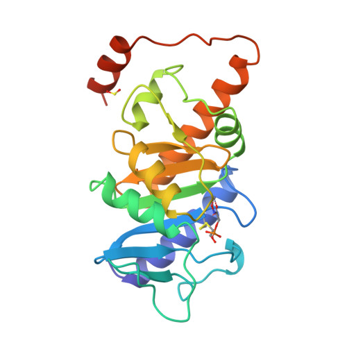
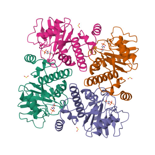
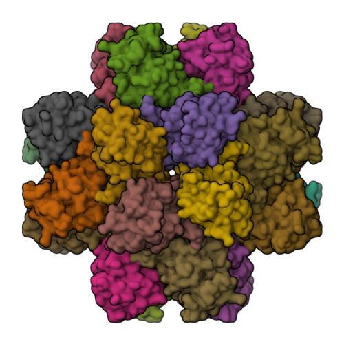
Catalytic mechanism of the metal-dependent fuculose aldolase from Escherichia coli as derived from the structure.

Dreyer, M.K.Schulz, G.E.

(1996) J Mol Biology 259: 458-466

  • DOI: https://doi.org/10.1006/jmbi.1996.0332
  • Primary Citation of Related Structures:  
    3FUA, 4FUA

  • PubMed Abstract: 

    The structure of L-fuculose-1-phosphate aldolase in a cubic crystal form has been determined with and without the inhibitor phosphoglycolohydroxamate at 2.4 and 2.7 angstrom (1 angstrom = 0.1 nm) resolution, respectively. This inhibitor mimics the enediolate transition state of the substrate moiety dihydroxyacetone phosphate. The structures showed that dihydroxyacetone phosphate ligates the zinc ion of this metal-dependent class II aldolase with its hydroxyl and keto oxygen atoms, shifting Glu73 away from the zinc coordination sphere to a non-polar environment. At this position Glu73 accepts a proton in the initial reaction step, producing the enediolate which is then stabilized by the zinc ion. The other substrate moiety L-lactaldehyde was modeled, because no binding structure is yet available.


  • Organizational Affiliation

    Institut für Organische Chemie und Biochemie, Albert-Ludwigs-Universität, Freiburg im Breisgau, Germany.


Macromolecules
Find similar proteins by:  (by identity cutoff)  |  3D Structure
Entity ID: 1
MoleculeChains Sequence LengthOrganismDetailsImage
L-FUCULOSE-1-PHOSPHATE ALDOLASE215Escherichia coliMutation(s): 0 
EC: 4.1.2.17
UniProt
Find proteins for P0AB87 (Escherichia coli (strain K12))
Explore P0AB87 
Go to UniProtKB:  P0AB87
Entity Groups  
Sequence Clusters30% Identity50% Identity70% Identity90% Identity95% Identity100% Identity
UniProt GroupP0AB87
Sequence Annotations
Expand
  • Reference Sequence
Experimental Data & Validation

Experimental Data

  • Method: X-RAY DIFFRACTION
  • Resolution: 2.43 Å
  • R-Value Work:  0.185 (Depositor), 0.170 (DCC) 
  • R-Value Observed: 0.185 (Depositor) 
Space Group: F 4 3 2
Unit Cell:
Length ( Å )Angle ( ˚ )
a = 183.6α = 90
b = 183.6β = 90
c = 183.6γ = 90
Software Package:
Software NamePurpose
X-PLORmodel building
X-PLORrefinement
XDSdata reduction
X-PLORphasing

Structure Validation

View Full Validation Report


Ligand Structure Quality Assessment 

Created with Raphaël 2.3.0Worse 01 BetterLigand structure goodness of fit to experimental dataBest fitted PGHClick on this verticalbar to view details

Entry History 

Deposition Data

Revision History  (Full details and data files)

  • Version 1.0: 1996-10-14
    Type: Initial release
  • Version 1.1: 2008-03-25
    Changes: Version format compliance
  • Version 1.2: 2011-07-13
    Changes: Derived calculations, Version format compliance
  • Version 2.0: 2024-06-05
    Changes: Atomic model, Data collection, Database references, Derived calculations, Other