4JF4

OXA-23 meropenem complex


Experimental Data Snapshot

  • Method: X-RAY DIFFRACTION
  • Resolution: 2.14 Å
  • R-Value Free: 
    0.254 (Depositor), 0.254 (DCC) 
  • R-Value Work: 
    0.199 (Depositor), 0.201 (DCC) 
  • R-Value Observed: 
    0.202 (Depositor) 

wwPDB Validation   3D Report Full Report


Ligand Structure Quality Assessment 


This is version 1.2 of the entry. See complete history


Literature

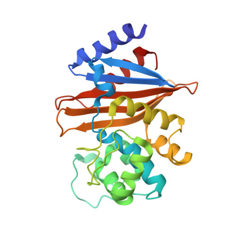
Structural Basis for Carbapenemase Activity of the OXA-23 beta-Lactamase from Acinetobacter baumannii.

Smith, C.A.Antunes, N.T.Stewart, N.K.Toth, M.Kumarasiri, M.Chang, M.Mobashery, S.Vakulenko, S.B.

(2013) Chem Biol 20: 1107-1115

  • DOI: https://doi.org/10.1016/j.chembiol.2013.07.015
  • Primary Citation of Related Structures:  
    4JF4, 4JF5, 4JF6

  • PubMed Abstract: 

    Dissemination of Acinetobacter baumannii strains harboring class D β-lactamases producing resistance to carbapenem antibiotics severely limits our ability to treat deadly Acinetobacter infections. Susceptibility determination in the A. baumannii background and kinetic studies with a homogeneous preparation of OXA-23 β-lactamase, the major carbapenemase present in A. baumannii, document the ability of this enzyme to manifest resistance to last-resort carbapenem antibiotics. We also report three X-ray structures of OXA-23: apo OXA-23 at two different pH values, and wild-type OXA-23 in complex with meropenem, a carbapenem substrate. The structures and dynamics simulations reveal an important role for Leu166, whose motion regulates the access of a hydrolytic water molecule to the acyl-enzyme species in imparting carbapenemase activity.


  • Organizational Affiliation

    Stanford Synchrotron Radiation Lightsource, Stanford University, Menlo Park, CA 94025, USA. Electronic address: csmith@slac.stanford.edu.


Macromolecules
Find similar proteins by:  (by identity cutoff)  |  3D Structure
Entity ID: 1
MoleculeChains Sequence LengthOrganismDetailsImage
Beta-lactamase
A, B
243Acinetobacter baumanniiMutation(s): 0 
Gene Names: ari-1bla(OXA-23)bla-OXA-23blaOXA-23OXA-23oxa23
EC: 3.5.2.6
UniProt
Find proteins for Q9L4P2 (Acinetobacter baumannii)
Explore Q9L4P2 
Go to UniProtKB:  Q9L4P2
Entity Groups  
Sequence Clusters30% Identity50% Identity70% Identity90% Identity95% Identity100% Identity
UniProt GroupQ9L4P2
Sequence Annotations
Expand
  • Reference Sequence
Small Molecules
Ligands 2 Unique
IDChains Name / Formula / InChI Key2D Diagram3D Interactions
MER
Query on MER

Download Ideal Coordinates CCD File 
D [auth A],
E [auth B]
(4R,5S)-3-{[(3S,5S)-5-(dimethylcarbamoyl)pyrrolidin-3-yl]sulfanyl}-5-[(2S,3R)-3-hydroxy-1-oxobutan-2-yl]-4-methyl-4,5-d ihydro-1H-pyrrole-2-carboxylic acid
C17 H27 N3 O5 S
DYQHXZPAIVAJRU-HTXLXMOSSA-N
EDO
Query on EDO

Download Ideal Coordinates CCD File 
C [auth A]1,2-ETHANEDIOL
C2 H6 O2
LYCAIKOWRPUZTN-UHFFFAOYSA-N
Experimental Data & Validation

Experimental Data

  • Method: X-RAY DIFFRACTION
  • Resolution: 2.14 Å
  • R-Value Free:  0.254 (Depositor), 0.254 (DCC) 
  • R-Value Work:  0.199 (Depositor), 0.201 (DCC) 
  • R-Value Observed: 0.202 (Depositor) 
Space Group: P 1 21 1
Unit Cell:
Length ( Å )Angle ( ˚ )
a = 49.467α = 90
b = 44.533β = 94.93
c = 131.367γ = 90
Software Package:
Software NamePurpose
Blu-Icedata collection
MOLREPphasing
PHENIXrefinement
XDSdata reduction
SCALAdata scaling

Structure Validation

View Full Validation Report


Ligand Structure Quality Assessment 


Entry History 

Deposition Data

Revision History  (Full details and data files)

  • Version 1.0: 2013-09-25
    Type: Initial release
  • Version 1.1: 2013-10-09
    Changes: Database references
  • Version 1.2: 2024-10-09
    Changes: Data collection, Database references, Derived calculations, Structure summary