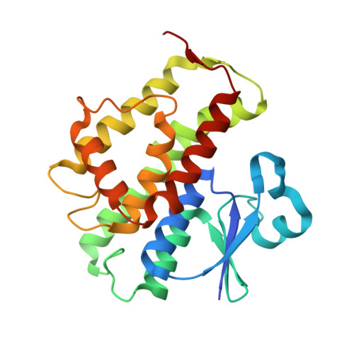
Comparison of epsilon- and delta-class glutathione S-transferases: the crystal structures of the glutathione S-transferases DmGSTE6 and DmGSTE7 from Drosophila melanogaster.
Scian, M., Le Trong, I., Mazari, A.M., Mannervik, B., Atkins, W.M., Stenkamp, R.E.(2015) Acta Crystallogr D Biol Crystallogr 71: 2089-2098
- PubMed: 26457432
- DOI: https://doi.org/10.1107/S1399004715013929
- Primary Citation of Related Structures:
4PNF, 4PNG - PubMed Abstract:
Cytosolic glutathione transferases (GSTs) comprise a large family of enzymes with canonical structures that diverge functionally and structurally among mammals, invertebrates and plants. Whereas mammalian GSTs have been characterized extensively with regard to their structure and function, invertebrate GSTs remain relatively unstudied. The invertebrate GSTs do, however, represent potentially important drug targets for infectious diseases and agricultural applications. In addition, it is essential to fully understand the structure and function of invertebrate GSTs, which play important roles in basic biological processes. Invertebrates harbor delta- and epsilon-class GSTs, which are not found in other organisms. Drosophila melanogaster GSTs (DmGSTs) are likely to contribute to detoxication or antioxidative stress during development, but they have not been fully characterized. Here, the structures of two epsilon-class GSTs from Drosophila, DmGSTE6 and DmGSTE7, are reported at 2.1 and 1.5 Å resolution, respectively, and are compared with other GSTs to identify structural features that might correlate with their biological functions. The structures of DmGSTE6 and DmGSTE7 are remarkably similar; the structures do not reveal obvious sources of the minor functional differences that have been observed. The main structural difference between the epsilon- and delta-class GSTs is the longer helix (A8) at the C-termini of the epsilon-class enzymes.
Organizational Affiliation:
Department of Medicinal Chemistry, University of Washington, Box 357610, Seattle, WA 98195-7610, USA.


