4R0O

Crystal structure of PEGylated plastocyanin at 4.2 A resolution


Experimental Data Snapshot

  • Method: X-RAY DIFFRACTION
  • Resolution: 4.20 Å
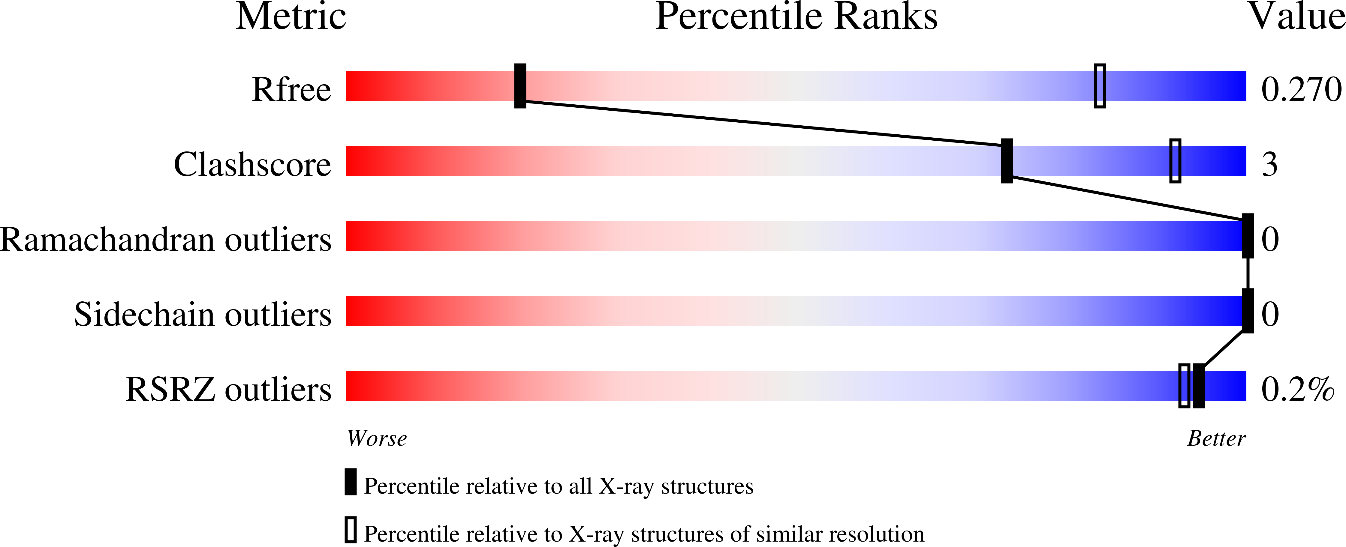
  • R-Value Free: 0.270 
  • R-Value Work: 0.229 
  • R-Value Observed: 0.230 

Starting Model: experimental
View more details

wwPDB Validation   3D Report Full Report


This is version 1.2 of the entry. See complete history


Literature

Structure of a PEGylated protein reveals a highly porous double-helical assembly.

Cattani, G.Vogeley, L.Crowley, P.B.

(2015) Nat Chem 7: 823-828

  • DOI: https://doi.org/10.1038/nchem.2342
  • Primary Citation of Related Structures:  
    4R0O

  • PubMed Abstract: 

    PEGylated proteins are a mainstay of the biopharmaceutical industry. Although the use of poly(ethylene glycol) (PEG) to increase particle size, stability and solubility is well-established, questions remain as to the structure of PEG-protein conjugates. Here we report the structural characterization of a model β-sheet protein (plastocyanin, 11.5 kDa) modified with a single PEG 5,000. An NMR spectroscopy study of the PEGylated conjugate indicated that the protein and PEG behaved as independent domains. A crystal structure revealed an extraordinary double-helical assembly of the conjugate, with the helices arranged orthogonally to yield a highly porous architecture. Electron density was not observed for the PEG chain, which indicates that it was disordered. The volume available per PEG chain in the crystal was within 10% of the calculated random coil volume. Together, these data support a minimal interaction between the protein and the synthetic polymer. Our work provides new possibilities for understanding this important class of protein-polymer hybrids and suggests a novel approach to engineering protein assemblies.


  • Organizational Affiliation

    School of Chemistry, National University of Ireland Galway, University Road, Galway, Ireland.


Macromolecules
Find similar proteins by:  (by identity cutoff)  |  3D Structure
Entity ID: 1
MoleculeChains Sequence LengthOrganismDetailsImage
Plastocyanin
A, B, C, D
106Leptolyngbya laminosaMutation(s): 0 
Gene Names: petE
UniProt
Find proteins for Q51883 (Phormidium laminosum)
Explore Q51883 
Go to UniProtKB:  Q51883
Entity Groups  
Sequence Clusters30% Identity50% Identity70% Identity90% Identity95% Identity100% Identity
UniProt GroupQ51883
Sequence Annotations
Expand
  • Reference Sequence
Small Molecules
Ligands 2 Unique
IDChains Name / Formula / InChI Key2D Diagram3D Interactions
LCY
Query on LCY

Download Ideal Coordinates CCD File 
F [auth A],
H [auth B],
J [auth C],
L [auth D]
1-methylpyrrolidine-2,5-dione
C5 H7 N O2
KYEACNNYFNZCST-UHFFFAOYSA-N
CU
Query on CU

Download Ideal Coordinates CCD File 
E [auth A],
G [auth B],
I [auth C],
K [auth D]
COPPER (II) ION
Cu
JPVYNHNXODAKFH-UHFFFAOYSA-N
Experimental Data & Validation

Experimental Data

  • Method: X-RAY DIFFRACTION
  • Resolution: 4.20 Å
  • R-Value Free: 0.270 
  • R-Value Work: 0.229 
  • R-Value Observed: 0.230 
  • Space Group: P 41 21 2
Unit Cell:
Length ( Å )Angle ( ˚ )
a = 116.14α = 90
b = 116.14β = 90
c = 172.14γ = 90
Software Package:
Software NamePurpose
PHASERphasing
PHENIXrefinement
XDSdata reduction
XSCALEdata scaling

Structure Validation

View Full Validation Report


Entry History 

Deposition Data

Revision History  (Full details and data files)

  • Version 1.0: 2015-10-07
    Type: Initial release
  • Version 1.1: 2023-09-20
    Changes: Data collection, Database references, Derived calculations, Refinement description
  • Version 1.2: 2024-11-06
    Changes: Structure summary