4W93 | pdb_00004w93

Human pancreatic alpha-amylase in complex with montbretin A


Experimental Data Snapshot

  • Method: X-RAY DIFFRACTION
  • Resolution: 1.35 Å
  • R-Value Free: 
    0.211 (Depositor), 0.210 (DCC) 
  • R-Value Work: 
    0.198 (Depositor), 0.200 (DCC) 
  • R-Value Observed: 
    0.198 (Depositor) 

wwPDB Validation   3D Report Full Report


Ligand Structure Quality Assessment 

Created with Raphaël 2.3.0Worse 01 BetterLigand structure goodness of fit to experimental dataBest fitted 3L9Click on this verticalbar to view details

This is version 2.3 of the entry. See complete history

Re-refinement Note

A newer entry is available that reflects an alternative modeling of the original data: 6OBX 6OCN


Literature

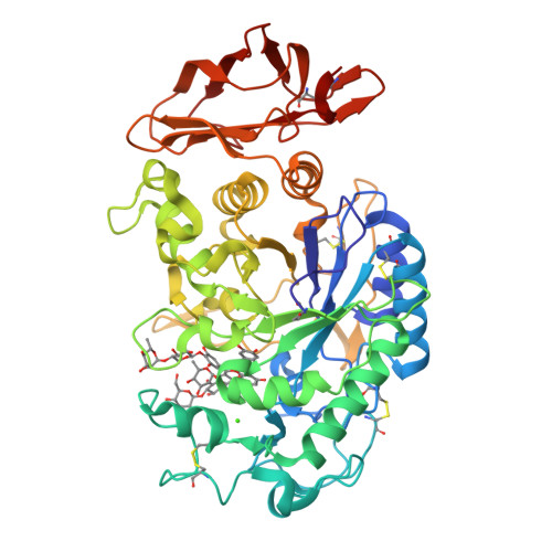
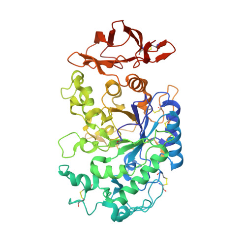
The amylase inhibitor montbretin A reveals a new glycosidase inhibition motif.

Williams, L.K.Zhang, X.Caner, S.Tysoe, C.Nguyen, N.T.Wicki, J.Williams, D.E.Coleman, J.McNeill, J.H.Yuen, V.Andersen, R.J.Withers, S.G.Brayer, G.D.

(2015) Nat Chem Biol 11: 691-696

  • DOI: https://doi.org/10.1038/nchembio.1865
  • Primary Citation of Related Structures:  
    4W93

  • PubMed Abstract: 

    The complex plant flavonol glycoside montbretin A is a potent (Ki = 8 nM) and specific inhibitor of human pancreatic α-amylase with potential as a therapeutic for diabetes and obesity. Controlled degradation studies on montbretin A, coupled with inhibition analyses, identified an essential high-affinity core structure comprising the myricetin and caffeic acid moieties linked via a disaccharide. X-ray structural analyses of the montbretin A-human α-amylase complex confirmed the importance of this core structure and revealed a novel mode of glycosidase inhibition wherein internal π-stacking interactions between the myricetin and caffeic acid organize their ring hydroxyls for optimal hydrogen bonding to the α-amylase catalytic residues D197 and E233. This novel inhibitory motif can be reproduced in a greatly simplified analog, offering potential for new strategies for glycosidase inhibition and therapeutic development.


  • Organizational Affiliation

    Department of Biochemistry and Molecular Biology, Faculty of Medicine, University of British Columbia, Vancouver, British Columbia, Canada.


Macromolecules
Find similar proteins by:  (by identity cutoff)  |  3D Structure
Entity ID: 1
MoleculeChains Sequence LengthOrganismDetailsImage
Pancreatic alpha-amylase496Homo sapiensMutation(s): 0 
Gene Names: AMY2A
EC: 3.2.1.1
UniProt & NIH Common Fund Data Resources
Find proteins for P04746 (Homo sapiens)
Explore P04746 
Go to UniProtKB:  P04746
PHAROS:  P04746
GTEx:  ENSG00000243480 
Entity Groups  
Sequence Clusters30% Identity50% Identity70% Identity90% Identity95% Identity100% Identity
UniProt GroupP04746
Sequence Annotations
Expand
  • Reference Sequence
Experimental Data & Validation

Experimental Data

  • Method: X-RAY DIFFRACTION
  • Resolution: 1.35 Å
  • R-Value Free:  0.211 (Depositor), 0.210 (DCC) 
  • R-Value Work:  0.198 (Depositor), 0.200 (DCC) 
  • R-Value Observed: 0.198 (Depositor) 
Space Group: P 21 21 21
Unit Cell:
Length ( Å )Angle ( ˚ )
a = 52.37α = 90
b = 74.1β = 90
c = 135.88γ = 90
Software Package:
Software NamePurpose
PHENIXrefinement

Structure Validation

View Full Validation Report


Ligand Structure Quality Assessment 

Created with Raphaël 2.3.0Worse 01 BetterLigand structure goodness of fit to experimental dataBest fitted 3L9Click on this verticalbar to view details

Entry History & Funding Information

Deposition Data


Funding OrganizationLocationGrant Number
Canadian Institutes of Health Research (CIHR)Canada--

Revision History  (Full details and data files)

  • Version 1.0: 2015-07-15
    Type: Initial release
  • Version 1.1: 2015-08-12
    Changes: Database references
  • Version 1.2: 2015-09-02
    Changes: Database references
  • Version 1.3: 2017-08-23
    Changes: Data collection, Derived calculations, Refinement description
  • Version 1.4: 2017-09-06
    Changes: Author supporting evidence
  • Version 2.0: 2019-12-25
    Changes: Database references, Polymer sequence
  • Version 2.1: 2020-01-08
    Changes: Author supporting evidence
  • Version 2.2: 2023-12-27
    Changes: Data collection, Database references, Derived calculations
  • Version 2.3: 2024-11-06
    Changes: Structure summary