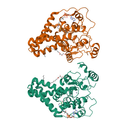
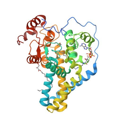
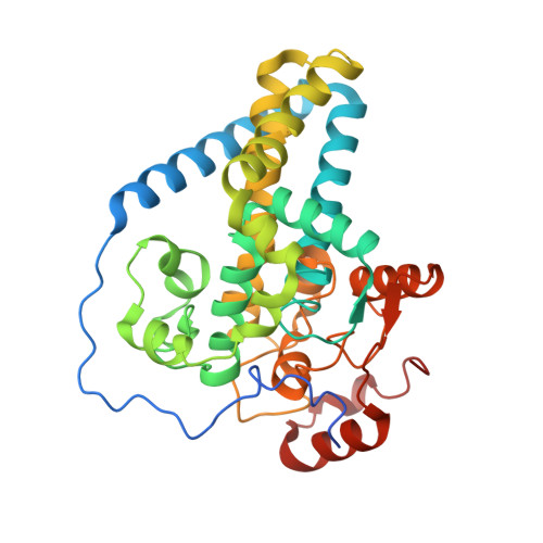
X-ray structure of a mammalian stearoyl-CoA desaturase.
Bai, Y., McCoy, J.G., Levin, E.J., Sobrado, P., Rajashankar, K.R., Fox, B.G., Zhou, M.(2015) Nature 524: 252-256
- PubMed: 26098370
- DOI: https://doi.org/10.1038/nature14549
- Primary Citation of Related Structures:
4YMK - PubMed Abstract:
Stearoyl-CoA desaturase (SCD) is conserved in all eukaryotes and introduces the first double bond into saturated fatty acyl-CoAs. Because the monounsaturated products of SCD are key precursors of membrane phospholipids, cholesterol esters and triglycerides, SCD is pivotal in fatty acid metabolism. Humans have two SCD homologues (SCD1 and SCD5), while mice have four (SCD1-SCD4). SCD1-deficient mice do not become obese or diabetic when fed a high-fat diet because of improved lipid metabolic profiles and insulin sensitivity. Thus, SCD1 is a pharmacological target in the treatment of obesity, diabetes and other metabolic diseases. SCD1 is an integral membrane protein located in the endoplasmic reticulum, and catalyses the formation of a cis-double bond between the ninth and tenth carbons of stearoyl- or palmitoyl-CoA. The reaction requires molecular oxygen, which is activated by a di-iron centre, and cytochrome b5, which regenerates the di-iron centre. To understand better the structural basis of these characteristics of SCD function, here we crystallize and solve the structure of mouse SCD1 bound to stearoyl-CoA at 2.6 Å resolution. The structure shows a novel fold comprising four transmembrane helices capped by a cytosolic domain, and a plausible pathway for lateral substrate access and product egress. The acyl chain of the bound stearoyl-CoA is enclosed in a tunnel buried in the cytosolic domain, and the geometry of the tunnel and the conformation of the bound acyl chain provide a structural basis for the regioselectivity and stereospecificity of the desaturation reaction. The dimetal centre is coordinated by a unique spacial arrangement of nine conserved histidine residues that implies a potentially novel mechanism for oxygen activation. The structure also illustrates a possible route for electron transfer from cytochrome b5 to the di-iron centre.
Organizational Affiliation:
Verna and Marrs McLean Department of Biochemistry and Molecular Biology, Baylor College of Medicine, Houston, Texas 77030, USA.


