5E95

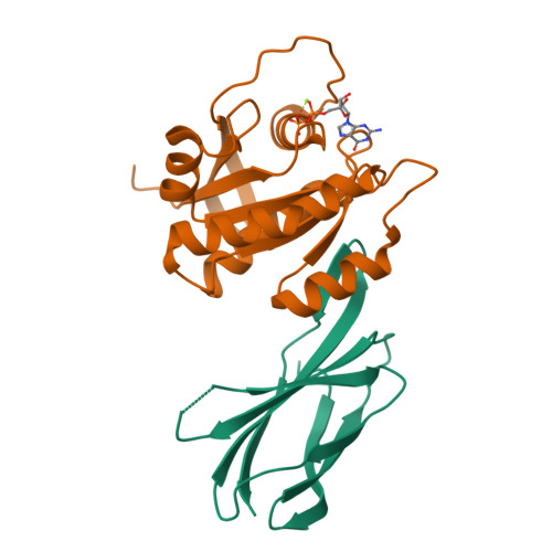
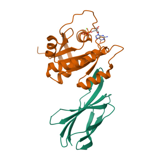
Crystal Structure of Mb(NS1)/H-Ras Complex


Experimental Data Snapshot

  • Method: X-RAY DIFFRACTION
  • Resolution: 1.40 Å
  • R-Value Free: 0.192 
  • R-Value Work: 0.163 
  • R-Value Observed: 0.164 

Starting Model: experimental
View more details

wwPDB Validation   3D Report Full Report


Ligand Structure Quality Assessment 


This is version 1.5 of the entry. See complete history


Literature

Inhibition of RAS function through targeting an allosteric regulatory site.

Spencer-Smith, R.Koide, A.Zhou, Y.Eguchi, R.R.Sha, F.Gajwani, P.Santana, D.Gupta, A.Jacobs, M.Herrero-Garcia, E.Cobbert, J.Lavoie, H.Smith, M.Rajakulendran, T.Dowdell, E.Okur, M.N.Dementieva, I.Sicheri, F.Therrien, M.Hancock, J.F.Ikura, M.Koide, S.O'Bryan, J.P.

(2017) Nat Chem Biol 13: 62-68

  • DOI: https://doi.org/10.1038/nchembio.2231
  • Primary Citation of Related Structures:  
    5E95

  • PubMed Abstract: 

    RAS GTPases are important mediators of oncogenesis in humans. However, pharmacological inhibition of RAS has proved challenging. Here we describe a functionally critical region, located outside the effector lobe of RAS, that can be targeted for inhibition. We developed NS1, a synthetic binding protein (monobody) that bound with high affinity to both GTP- and GDP-bound states of H-RAS and K-RAS but not N-RAS. NS1 potently inhibited growth factor signaling and oncogenic H-RAS- and K-RAS-mediated signaling and transformation but did not block oncogenic N-RAS, BRAF or MEK1. NS1 bound the α4-β6-α5 region of RAS, which disrupted RAS dimerization and nanoclustering and led to blocking of CRAF-BRAF heterodimerization and activation. These results establish the importance of the α4-β6-α5 interface in RAS-mediated signaling and define a previously unrecognized site in RAS for inhibiting RAS function.


  • Organizational Affiliation

    Department of Pharmacology, University of Illinois at Chicago, Chicago, Illinois, USA.


Macromolecules
Find similar proteins by:  (by identity cutoff)  |  3D Structure
Entity ID: 1
MoleculeChains Sequence LengthOrganismDetailsImage
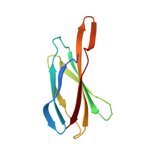
Mb(NS1)A [auth B]97Homo sapiensMutation(s): 0 
Sequence Annotations
Expand
  • Reference Sequence
Find similar proteins by:  (by identity cutoff)  |  3D Structure
Entity ID: 2
MoleculeChains Sequence LengthOrganismDetailsImage
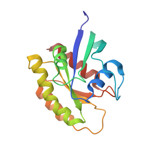
GTPase HRasB [auth A]168Homo sapiensMutation(s): 0 
Gene Names: HRASHRAS1
UniProt & NIH Common Fund Data Resources
Find proteins for P01112 (Homo sapiens)
Go to UniProtKB:  P01112
PHAROS:  P01112
GTEx:  ENSG00000174775 
Sequence Annotations
Expand
  • Reference Sequence
Small Molecules
Ligands 2 Unique
IDChains Name / Formula / InChI Key2D Diagram3D Interactions
GDP
Query on GDP

Download Ideal Coordinates CCD File 
C [auth A]GUANOSINE-5'-DIPHOSPHATE
C10 H15 N5 O11 P2
QGWNDRXFNXRZMB-UUOKFMHZSA-N
MG
Query on MG

Download Ideal Coordinates CCD File 
D [auth A]MAGNESIUM ION
Mg
JLVVSXFLKOJNIY-UHFFFAOYSA-N
Experimental Data & Validation

Experimental Data

  • Method: X-RAY DIFFRACTION
  • Resolution: 1.40 Å
  • R-Value Free: 0.192 
  • R-Value Work: 0.163 
  • R-Value Observed: 0.164 
  • Space Group: P 1
Unit Cell:
Length ( Å )Angle ( ˚ )
a = 35.897α = 86.3
b = 36.276β = 74.23
c = 54.348γ = 85.2
Software Package:
Software NamePurpose
PHENIXrefinement
Cootmodel building
HKL-3000data scaling
HKL-3000data reduction
PHASERphasing

Structure Validation

View Full Validation Report


Ligand Structure Quality Assessment 


Entry History & Funding Information

Deposition Data


Funding OrganizationLocationGrant Number
National Institutes of Health/National Institute of General Medical Sciences (NIH/NIGMS)United StatesR01-GM090324
Department of Energy (DOE, United States)United StatesDE-AC02-06CH11357
The Chicago Biomedical Consortium with support from The Searle Funds at The Chicago Community TrustUnited States--

Revision History  (Full details and data files)

  • Version 1.0: 2016-11-02
    Type: Initial release
  • Version 1.1: 2016-11-23
    Changes: Database references
  • Version 1.2: 2016-12-28
    Changes: Database references
  • Version 1.3: 2017-09-27
    Changes: Author supporting evidence
  • Version 1.4: 2019-12-04
    Changes: Author supporting evidence
  • Version 1.5: 2023-09-27
    Changes: Data collection, Database references, Refinement description