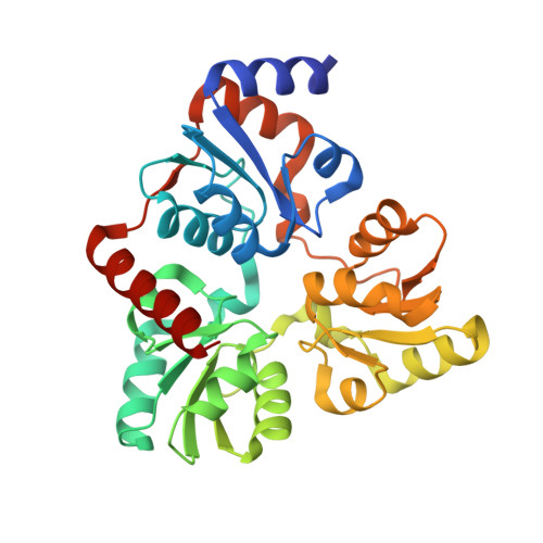
5KTT

Crystal structure of Pyrococcus horikoshii quinolinate synthase (NadA) with bound L-malate and Fe4S4 cluster


Experimental Data Snapshot

  • Method: X-RAY DIFFRACTION
  • Resolution: 1.50 Å
  • R-Value Free: 0.193 
  • R-Value Work: 0.146 
  • R-Value Observed: 0.148 

Starting Model: experimental
View more details

wwPDB Validation   3D Report Full Report


This is version 1.5 of the entry. See complete history


Literature

Crystal Structures of the Iron-Sulfur Cluster-Dependent Quinolinate Synthase in Complex with Dihydroxyacetone Phosphate, Iminoaspartate Analogues, and Quinolinate.

Fenwick, M.K.Ealick, S.E.

(2016) Biochemistry 55: 4135-4139

  • DOI: https://doi.org/10.1021/acs.biochem.6b00626
  • Primary Citation of Related Structures:  
    5KTM, 5KTN, 5KTO, 5KTP, 5KTR, 5KTS, 5KTT

  • PubMed Abstract: 

    The quinolinate synthase of prokaryotes and photosynthetic eukaryotes, NadA, contains a [4Fe-4S] cluster with unknown function. We report crystal structures of Pyrococcus horikoshii NadA in complex with dihydroxyacetone phosphate (DHAP), iminoaspartate analogues, and quinolinate. DHAP adopts a nearly planar conformation and chelates the [4Fe-4S] cluster via its keto and hydroxyl groups. The active site architecture suggests that the cluster acts as a Lewis acid in enediolate formation, like zinc in class II aldolases. The DHAP and putative iminoaspartate structures suggest a model for a condensed intermediate. The ensemble of structures suggests a two-state system, which may be exploited in early steps.


  • Organizational Affiliation

    Department of Chemistry and Chemical Biology, Cornell University , Ithaca, New York 14853, United States.


Macromolecules
Find similar proteins by:  (by identity cutoff)  |  3D Structure
Entity ID: 1
MoleculeChains Sequence LengthOrganismDetailsImage
Quinolinate synthase A304Pyrococcus horikoshii OT3Mutation(s): 0 
Gene Names: nadAPH0013
EC: 2.5.1.72
UniProt
Find proteins for O57767 (Pyrococcus horikoshii (strain ATCC 700860 / DSM 12428 / JCM 9974 / NBRC 100139 / OT-3))
Explore O57767 
Go to UniProtKB:  O57767
Entity Groups  
Sequence Clusters30% Identity50% Identity70% Identity90% Identity95% Identity100% Identity
UniProt GroupO57767
Sequence Annotations
Expand
  • Reference Sequence
Experimental Data & Validation

Experimental Data

  • Method: X-RAY DIFFRACTION
  • Resolution: 1.50 Å
  • R-Value Free: 0.193 
  • R-Value Work: 0.146 
  • R-Value Observed: 0.148 
  • Space Group: P 1 21 1
Unit Cell:
Length ( Å )Angle ( ˚ )
a = 47.333α = 90
b = 54.639β = 90.79
c = 55.613γ = 90
Software Package:
Software NamePurpose
PHENIXrefinement
PHENIXphasing
HKL-2000data reduction
HKL-2000data scaling

Structure Validation

View Full Validation Report


Entry History & Funding Information

Deposition Data


Funding OrganizationLocationGrant Number
National Institutes of Health/National Institute of General Medical Sciences (NIH/NIGMS)United StatesGM103403
Department of Energy (DOE, United States)United StatesDE-AC02-06CH11357
National Institutes of Health/National Institute of General Medical Sciences (NIH/NIGMS)United StatesGM103485

Revision History  (Full details and data files)

  • Version 1.0: 2016-07-27
    Type: Initial release
  • Version 1.1: 2016-08-10
    Changes: Database references
  • Version 1.2: 2016-08-17
    Changes: Database references
  • Version 1.3: 2017-09-20
    Changes: Author supporting evidence, Database references, Derived calculations
  • Version 1.4: 2019-12-04
    Changes: Author supporting evidence
  • Version 1.5: 2023-10-04
    Changes: Data collection, Database references, Refinement description