5LFT | pdb_00005lft

Crystal structure of cytochrome c - Bromo-trisulfonatocalix[4]arene complexes


Experimental Data Snapshot

  • Method: X-RAY DIFFRACTION
  • Resolution: 1.25 Å
  • R-Value Free: 
    0.239 (Depositor), 0.240 (DCC) 
  • R-Value Work: 
    0.213 (Depositor), 0.210 (DCC) 
  • R-Value Observed: 
    0.215 (Depositor) 

Starting Model: experimental
View more details

wwPDB Validation   3D Report Full Report


Ligand Structure Quality Assessment 

Created with Raphaël 2.3.0Worse 01 BetterLigand structure goodness of fit to experimental dataBest fitted 6VBClick on this verticalbar to view detailsBest fitted HECClick on this verticalbar to view details

This is version 2.2 of the entry. See complete history


Literature

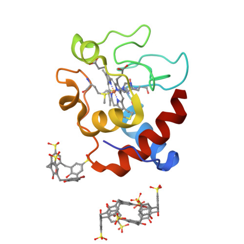
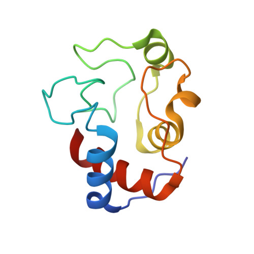
Protein Recognition by Functionalized Sulfonatocalix[4]arenes.

Doolan, A.M.Rennie, M.L.Crowley, P.B.

(2018) Chemistry 24: 984-991

  • DOI: https://doi.org/10.1002/chem.201704931
  • Primary Citation of Related Structures:  
    5KPF, 5LFT, 5T8W

  • PubMed Abstract: 

    The interactions of two mono-functionalized sulfonatocalix[4]arenes with cytochrome c were investigated by structural and thermodynamic methods. The replacement of a single sulfonate with either a bromo or a phenyl substituent resulted in altered recognition of cytochrome c as evidenced by X-ray crystallography. The bromo-substituted ligand yielded a new binding mode in which a self-encapsulated calixarene dimer contributed to crystal packing. This ligand also formed a weak halogen bond with the protein. The phenyl-substituted ligand was bound to Lys4 of cytochrome c, in a 1.7 Å resolution crystal structure. A dimeric packing arrangement mediated by ligand-ligand contacts in the crystal suggested a possible assembly mechanism. The different protein recognition properties of these calixarenes are discussed.


  • Organizational Affiliation

    School of Chemistry, National University of Ireland Galway, University Road, Galway, Ireland.


Macromolecules
Find similar proteins by:  (by identity cutoff)  |  3D Structure
Entity ID: 1
MoleculeChains Sequence LengthOrganismDetailsImage
Cytochrome c iso-1108Saccharomyces cerevisiaeMutation(s): 0 
Gene Names: CYC1YJR048WJ1653
UniProt
Find proteins for P00044 (Saccharomyces cerevisiae (strain ATCC 204508 / S288c))
Explore P00044 
Go to UniProtKB:  P00044
Entity Groups  
Sequence Clusters30% Identity50% Identity70% Identity90% Identity95% Identity100% Identity
UniProt GroupP00044
Sequence Annotations
Expand
  • Reference Sequence
Experimental Data & Validation

Experimental Data

  • Method: X-RAY DIFFRACTION
  • Resolution: 1.25 Å
  • R-Value Free:  0.239 (Depositor), 0.240 (DCC) 
  • R-Value Work:  0.213 (Depositor), 0.210 (DCC) 
  • R-Value Observed: 0.215 (Depositor) 
Space Group: P 2 21 21
Unit Cell:
Length ( Å )Angle ( ˚ )
a = 34.746α = 90
b = 38.045β = 90
c = 89.65γ = 90
Software Package:
Software NamePurpose
SCALAdata scaling
PHASERphasing
PHENIXrefinement
PDB_EXTRACTdata extraction
XDSdata reduction

Structure Validation

View Full Validation Report


Ligand Structure Quality Assessment 

Created with Raphaël 2.3.0Worse 01 BetterLigand structure goodness of fit to experimental dataBest fitted 6VBClick on this verticalbar to view detailsBest fitted HECClick on this verticalbar to view details

Entry History & Funding Information

Deposition Data


Funding OrganizationLocationGrant Number
Science Foundation IrelandIreland13/ERC/B2912

Revision History  (Full details and data files)

  • Version 1.0: 2017-07-12
    Type: Initial release
  • Version 1.1: 2018-02-28
    Changes: Database references
  • Version 2.0: 2019-01-23
    Changes: Atomic model, Data collection, Derived calculations, Non-polymer description, Structure summary
  • Version 2.1: 2024-01-10
    Changes: Data collection, Database references, Derived calculations, Refinement description
  • Version 2.2: 2024-10-23
    Changes: Structure summary