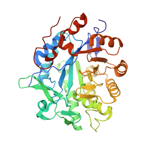
5LGX

Structure of Oxidised Pentaerythritol Tetranitrate Reductase


Experimental Data Snapshot

  • Method: X-RAY DIFFRACTION
  • Resolution: 1.50 Å
  • R-Value Free: 0.200 
  • R-Value Work: 0.156 
  • R-Value Observed: 0.158 

Starting Model: experimental
View more details

wwPDB Validation   3D Report Full Report


Ligand Structure Quality Assessment 


This is version 1.2 of the entry. See complete history


Literature

Combining X-ray and neutron crystallography with spectroscopy.

Kwon, H.Smith, O.Raven, E.L.Moody, P.C.

(2017) Acta Crystallogr D Struct Biol 73: 141-147

  • DOI: https://doi.org/10.1107/S2059798316016314
  • Primary Citation of Related Structures:  
    5LGX, 5LGZ

  • PubMed Abstract: 

    X-ray protein crystallography has, through the determination of the three-dimensional structures of enzymes and their complexes, been essential to the understanding of biological chemistry. However, as X-rays are scattered by electrons, the technique has difficulty locating the presence and position of H atoms (and cannot locate H + ions), knowledge of which is often crucially important for the understanding of enzyme mechanism. Furthermore, X-ray irradiation, through photoelectronic effects, will perturb the redox state in the crystal. By using single-crystal spectrophotometry, reactions taking place in the crystal can be monitored, either to trap intermediates or follow photoreduction during X-ray data collection. By using neutron crystallography, the positions of H atoms can be located, as it is the nuclei rather than the electrons that scatter neutrons, and the scattering length is not determined by the atomic number. Combining the two techniques allows much greater insight into both reaction mechanism and X-ray-induced photoreduction.


  • Organizational Affiliation

    Henry Wellcome Laboratories for Structural Biology, Leicester Institute for Structural and Chemical Biology and Department of Molecular and Cell Biology, University of Leicester, Lancaster Road, Leicester LE1 7RH, England.


Macromolecules
Find similar proteins by:  (by identity cutoff)  |  3D Structure
Entity ID: 1
MoleculeChains Sequence LengthOrganismDetailsImage
Pentaerythritol tetranitrate reductase363Enterobacter cloacaeMutation(s): 0 
Gene Names: onr
UniProt
Find proteins for P71278 (Enterobacter cloacae)
Explore P71278 
Go to UniProtKB:  P71278
Entity Groups  
Sequence Clusters30% Identity50% Identity70% Identity90% Identity95% Identity100% Identity
UniProt GroupP71278
Sequence Annotations
Expand
  • Reference Sequence
Experimental Data & Validation

Experimental Data

  • Method: X-RAY DIFFRACTION
  • Resolution: 1.50 Å
  • R-Value Free: 0.200 
  • R-Value Work: 0.156 
  • R-Value Observed: 0.158 
  • Space Group: P 21 21 21
Unit Cell:
Length ( Å )Angle ( ˚ )
a = 56.76α = 90
b = 68.587β = 90
c = 88.574γ = 90
Software Package:
Software NamePurpose
REFMACrefinement
iMOSFLMdata reduction
Aimlessdata scaling
PHASERphasing

Structure Validation

View Full Validation Report


Ligand Structure Quality Assessment 


Entry History 

Deposition Data

Revision History  (Full details and data files)

  • Version 1.0: 2017-02-15
    Type: Initial release
  • Version 1.1: 2017-03-01
    Changes: Database references
  • Version 1.2: 2024-01-10
    Changes: Data collection, Database references, Refinement description