5NTT

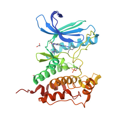
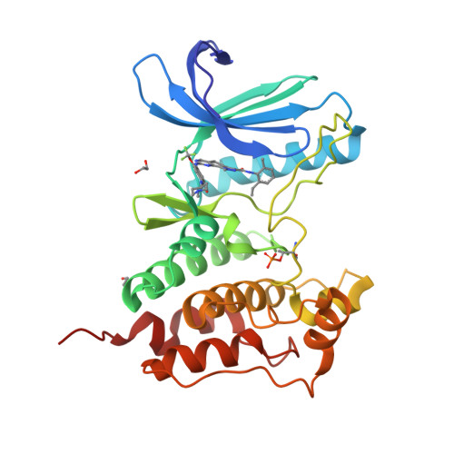
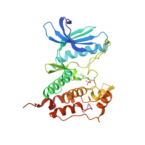
Crystal structure of human Mps1 (TTK) C604Y mutant in complex with NMS-P715


Experimental Data Snapshot

  • Method: X-RAY DIFFRACTION
  • Resolution: 2.75 Å
  • R-Value Free: 0.230 
  • R-Value Work: 0.199 
  • R-Value Observed: 0.201 

Starting Model: experimental
View more details

wwPDB Validation   3D Report Full Report


Ligand Structure Quality Assessment 


This is version 1.4 of the entry. See complete history


Literature

Understanding inhibitor resistance in Mps1 kinase through novel biophysical assays and structures.

Hiruma, Y.Koch, A.Hazraty, N.Tsakou, F.Medema, R.H.Joosten, R.P.Perrakis, A.

(2017) J Biol Chem 292: 14496-14504

  • DOI: https://doi.org/10.1074/jbc.M117.783555

  • PubMed Abstract: 

    Monopolar spindle 1 (Mps1/TTK) is a protein kinase essential in mitotic checkpoint signaling, preventing anaphase until all chromosomes are properly attached to spindle microtubules. Mps1 has emerged as a potential target for cancer therapy, and a variety of compounds have been developed to inhibit its kinase activity. Mutations in the catalytic domain of Mps1 that give rise to inhibitor resistance, but retain catalytic activity and do not display cross-resistance to other Mps1 inhibitors, have been described. Here we characterize the interactions of two such mutants, Mps1 C604Y and C604W, which raise resistance to two closely related compounds, NMS-P715 and its derivative Cpd-5, but not to the well characterized Mps1 inhibitor, reversine. We show that estimates of the IC 50 (employing a novel specific and efficient assay that utilizes a fluorescently labeled substrate) and the binding affinity ( K D ) indicate that, in both mutants, Cpd-5 should be better tolerated than the closely related NMS-P715. To gain further insight, we determined the crystal structure of the Mps1 kinase mutants bound to Cpd-5 and NMS-P715 and compared the binding modes of Cpd-5, NMS-P715, and reversine. The difference in steric hindrance between Tyr/Trp 604 and the trifluoromethoxy moiety of NMS-P715, the methoxy moiety of Cpd-5, and complete absence of such a group in reversine, account for differences we observe in vitro Our analysis enforces the notion that inhibitors targeting Mps1 drug-resistant mutations can emerge as a feasible intervention strategy based on existing scaffolds, if the clinical need arises.


  • Organizational Affiliation

    From the Divisions of Biochemistry and.


Macromolecules
Find similar proteins by:  (by identity cutoff)  |  3D Structure
Entity ID: 1
MoleculeChains Sequence LengthOrganismDetailsImage
Dual specificity protein kinase TTK287Homo sapiensMutation(s): 1 
Gene Names: TTKMPS1MPS1L1
EC: 2.7.12.1
UniProt & NIH Common Fund Data Resources
Find proteins for P33981 (Homo sapiens)
Explore P33981 
Go to UniProtKB:  P33981
PHAROS:  P33981
GTEx:  ENSG00000112742 
Entity Groups  
Sequence Clusters30% Identity50% Identity70% Identity90% Identity95% Identity100% Identity
UniProt GroupP33981
Sequence Annotations
Expand
  • Reference Sequence
Small Molecules
Ligands 2 Unique
IDChains Name / Formula / InChI Key2D Diagram3D Interactions
SVE
Query on SVE

Download Ideal Coordinates CCD File 
B [auth A]N-(2,6-DIETHYLPHENYL)-1-METHYL-8-({4-[(1-METHYLPIPERIDIN-4-YL)CARBAMOYL]-2-(TRIFLUOROMETHOXY)PHENYL}AMINO)-4,5-DIHYDRO-1H-PYRAZOLO[4,3-H]QUINAZOLINE-3-CARBOXAMIDE
C35 H39 F3 N8 O3
JFOAJUGFHDCBJJ-UHFFFAOYSA-N
EDO
Query on EDO

Download Ideal Coordinates CCD File 
C [auth A],
D [auth A]
1,2-ETHANEDIOL
C2 H6 O2
LYCAIKOWRPUZTN-UHFFFAOYSA-N
Modified Residues  1 Unique
IDChains TypeFormula2D DiagramParent
TPO
Query on TPO
A
L-PEPTIDE LINKINGC4 H10 N O6 PTHR
Experimental Data & Validation

Experimental Data

  • Method: X-RAY DIFFRACTION
  • Resolution: 2.75 Å
  • R-Value Free: 0.230 
  • R-Value Work: 0.199 
  • R-Value Observed: 0.201 
  • Space Group: I 2 2 2
Unit Cell:
Length ( Å )Angle ( ˚ )
a = 70.672α = 90
b = 112.01β = 90
c = 116.126γ = 90
Software Package:
Software NamePurpose
Aimlessdata scaling
PHASERphasing
REFMACrefinement
PDB_EXTRACTdata extraction
XDSdata reduction
PHASERphasing

Structure Validation

View Full Validation Report


Ligand Structure Quality Assessment 


Entry History & Funding Information

Deposition Data


Funding OrganizationLocationGrant Number
Netherlands Organization for Scientific ResearchNetherlands722.015.008
Netherlands Organization for Scientific ResearchNetherlands723.013.003
KWF Dutch Cancer SocietyNetherlands2012-5427

Revision History  (Full details and data files)

  • Version 1.0: 2017-07-26
    Type: Initial release
  • Version 1.1: 2017-08-02
    Changes: Database references
  • Version 1.2: 2017-09-13
    Changes: Database references
  • Version 1.3: 2024-01-17
    Changes: Data collection, Database references, Refinement description
  • Version 1.4: 2024-11-13
    Changes: Structure summary