In vitro and in vivo characterization of threeCellvibrio japonicusglycoside hydrolase family 5 members reveals potent xyloglucan backbone-cleaving functions.
Attia, M.A., Nelson, C.E., Offen, W.A., Jain, N., Davies, G.J., Gardner, J.G., Brumer, H.(2018) Biotechnol Biofuels 11: 45-45
- PubMed: 29467823
- DOI: https://doi.org/10.1186/s13068-018-1039-6
- Primary Citation of Related Structures:
5OYC, 5OYD, 5OYE - PubMed Abstract:
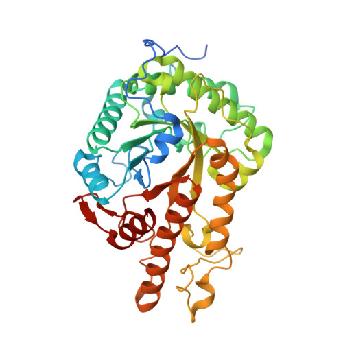
Xyloglucan (XyG) is a ubiquitous and fundamental polysaccharide of plant cell walls. Due to its structural complexity, XyG requires a combination of backbone-cleaving and sidechain-debranching enzymes for complete deconstruction into its component monosaccharides. The soil saprophyte Cellvibrio japonicus has emerged as a genetically tractable model system to study biomass saccharification, in part due to its innate capacity to utilize a wide range of plant polysaccharides for growth. Whereas the downstream debranching enzymes of the xyloglucan utilization system of C. japonicus have been functionally characterized, the requisite backbone-cleaving endo -xyloglucanases were unresolved. Combined bioinformatic and transcriptomic analyses implicated three glycoside hydrolase family 5 subfamily 4 (GH5_4) members, with distinct modular organization, as potential keystone endo -xyloglucanases in C. japonicus . Detailed biochemical and enzymatic characterization of the GH5_4 modules of all three recombinant proteins confirmed particularly high specificities for the XyG polysaccharide versus a panel of other cell wall glycans, including mixed-linkage beta-glucan and cellulose. Moreover, product analysis demonstrated that all three enzymes generated XyG oligosaccharides required for subsequent saccharification by known exo -glycosidases. Crystallographic analysis of GH5D, which was the only GH5_4 member specifically and highly upregulated during growth on XyG, in free, product-complex, and active-site affinity-labelled forms revealed the molecular basis for the exquisite XyG specificity among these GH5_4 enzymes. Strikingly, exhaustive reverse-genetic analysis of all three GH5_4 members and a previously biochemically characterized GH74 member failed to reveal a growth defect, thereby indicating functional compensation in vivo, both among members of this cohort and by other, yet unidentified, xyloglucanases in C. japonicus . Our systems-based analysis indicates distinct substrate-sensing (GH74, GH5E, GH5F) and attack-mounting (GH5D) functions for the endo -xyloglucanases characterized here. Through a multi-faceted, molecular systems-based approach, this study provides a new insight into the saccharification pathway of xyloglucan utilization system of C. japonicus . The detailed structural-functional characterization of three distinct GH5_4 endo -xyloglucanases will inform future bioinformatic predictions across species, and provides new CAZymes with defined specificity that may be harnessed in industrial and other biotechnological applications.
Organizational Affiliation:
1Michael Smith Laboratories, University of British Columbia, 2185 East Mall, Vancouver, BC V6T 1Z4 Canada.


