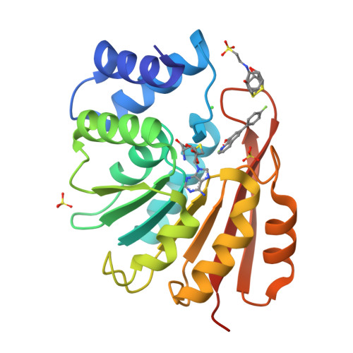
Crystal Structure of a COMT complex
Lerner, C., Jakob-Roetne, R., Groebke-Zbinden, K., Buettelmann, B., Rudolph, M.G.To be published.
Experimental Data Snapshot
Starting Model: other
View more details
Entity ID: 1 | |||||
|---|---|---|---|---|---|
| Molecule | Chains | Sequence Length | Organism | Details | Image |
| Catechol O-methyltransferase | 221 | Rattus norvegicus | Mutation(s): 0 Gene Names: Comt EC: 2.1.1.6 |  | |
UniProt | |||||
Find proteins for P22734 (Rattus norvegicus) Explore P22734 Go to UniProtKB: P22734 | |||||
Entity Groups | |||||
| Sequence Clusters | 30% Identity50% Identity70% Identity90% Identity95% Identity100% Identity | ||||
| UniProt Group | P22734 | ||||
Sequence AnnotationsExpand | |||||
| |||||
| Ligands 7 Unique | |||||
|---|---|---|---|---|---|
| ID | Chains | Name / Formula / InChI Key | 2D Diagram | 3D Interactions | |
| SAH Query on SAH | H [auth A] | S-ADENOSYL-L-HOMOCYSTEINE C14 H20 N6 O5 S ZJUKTBDSGOFHSH-WFMPWKQPSA-N |  | ||
| 7JS Query on 7JS | I [auth A] | 6-(4-fluorophenyl)quinolin-8-ol C15 H10 F N O FTIUFDNWKCYJNH-UHFFFAOYSA-N |  | ||
| NHE Query on NHE | G [auth A] | 2-[N-CYCLOHEXYLAMINO]ETHANE SULFONIC ACID C8 H17 N O3 S MKWKNSIESPFAQN-UHFFFAOYSA-N |  | ||
| D1D Query on D1D | F [auth A] | (4S,5S)-1,2-DITHIANE-4,5-DIOL C4 H8 O2 S2 YPGMOWHXEQDBBV-QWWZWVQMSA-N |  | ||
| SO4 Query on SO4 | D [auth A], E [auth A] | SULFATE ION O4 S QAOWNCQODCNURD-UHFFFAOYSA-L |  | ||
| CL Query on CL | C [auth A] | CHLORIDE ION Cl VEXZGXHMUGYJMC-UHFFFAOYSA-M |  | ||
| MG Query on MG | B [auth A] | MAGNESIUM ION Mg JLVVSXFLKOJNIY-UHFFFAOYSA-N |  | ||
| Length ( Å ) | Angle ( ˚ ) |
|---|---|
| a = 50.047 | α = 90 |
| b = 54.205 | β = 90 |
| c = 80.932 | γ = 90 |
| Software Name | Purpose |
|---|---|
| XSCALE | data scaling |
| REFMAC | refinement |
| PDB_EXTRACT | data extraction |
| XDS | data reduction |
| PHASER | phasing |
RCSB PDB is hosted by

RCSB PDB is a member of the