5UGH

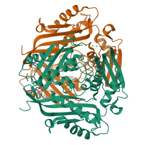
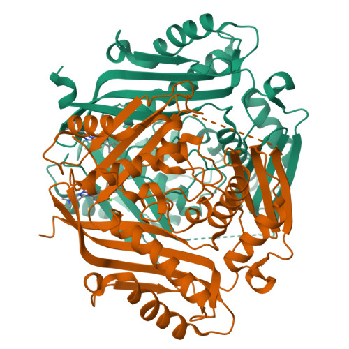
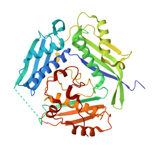
Crystal structure of Mat2a bound to the allosteric inhibitor PF-02929366


Experimental Data Snapshot

  • Method: X-RAY DIFFRACTION
  • Resolution: 2.06 Å
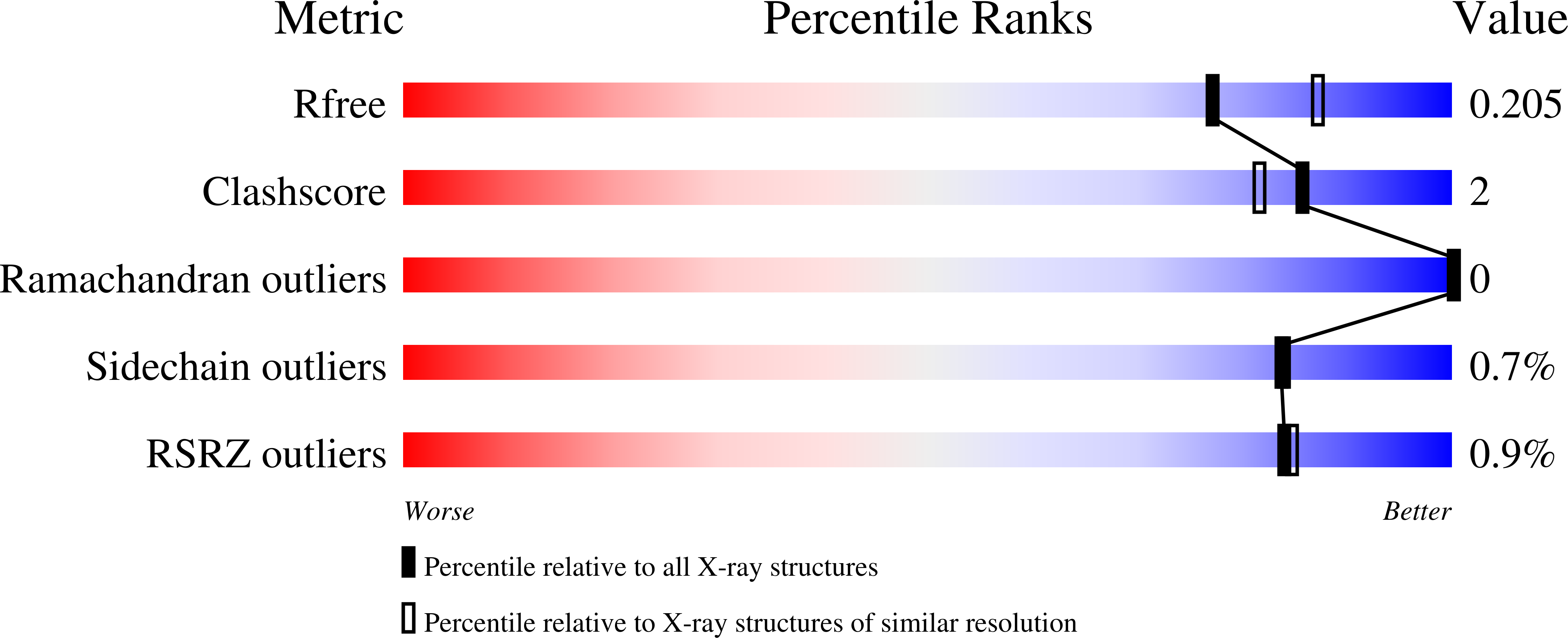
  • R-Value Free: 0.203 
  • R-Value Work: 0.176 
  • R-Value Observed: 0.177 

Starting Model: experimental
View more details

wwPDB Validation   3D Report Full Report


Ligand Structure Quality Assessment 


This is version 2.1 of the entry. See complete history


Literature

Targeting S-adenosylmethionine biosynthesis with a novel allosteric inhibitor of Mat2A.

Quinlan, C.L.Kaiser, S.E.Bolanos, B.Nowlin, D.Grantner, R.Karlicek-Bryant, S.Feng, J.L.Jenkinson, S.Freeman-Cook, K.Dann, S.G.Wang, X.Wells, P.A.Fantin, V.R.Stewart, A.E.Grant, S.K.

(2017) Nat Chem Biol 13: 785-792

  • DOI: https://doi.org/10.1038/nchembio.2384
  • Primary Citation of Related Structures:  
    5UGH

  • PubMed Abstract: 

    S-Adenosyl-L-methionine (SAM) is an enzyme cofactor used in methyl transfer reactions and polyamine biosynthesis. The biosynthesis of SAM from ATP and L-methionine is performed by the methionine adenosyltransferase enzyme family (Mat; EC 2.5.1.6). Human methionine adenosyltransferase 2A (Mat2A), the extrahepatic isoform, is often deregulated in cancer. We identified a Mat2A inhibitor, PF-9366, that binds an allosteric site on Mat2A that overlaps with the binding site for the Mat2A regulator, Mat2B. Studies exploiting PF-9366 suggested a general mode of Mat2A allosteric regulation. Allosteric binding of PF-9366 or Mat2B altered the Mat2A active site, resulting in increased substrate affinity and decreased enzyme turnover. These data support a model whereby Mat2B functions as an inhibitor of Mat2A activity when methionine or SAM levels are high, yet functions as an activator of Mat2A when methionine or SAM levels are low. The ramification of Mat2A activity modulation in cancer cells is also described.


  • Organizational Affiliation

    Oncology Research and Development, Pfizer Inc., San Diego, California, USA.


Macromolecules
Find similar proteins by:  (by identity cutoff)  |  3D Structure
Entity ID: 1
MoleculeChains Sequence LengthOrganismDetailsImage
S-adenosylmethionine synthase isoform type-2
A, B, C, D
397Homo sapiensMutation(s): 0 
Gene Names: MAT2AAMS2MATA2
EC: 2.5.1.6
UniProt & NIH Common Fund Data Resources
Find proteins for P31153 (Homo sapiens)
Explore P31153 
Go to UniProtKB:  P31153
PHAROS:  P31153
GTEx:  ENSG00000168906 
Entity Groups  
Sequence Clusters30% Identity50% Identity70% Identity90% Identity95% Identity100% Identity
UniProt GroupP31153
Sequence Annotations
Expand
  • Reference Sequence
Experimental Data & Validation

Experimental Data

  • Method: X-RAY DIFFRACTION
  • Resolution: 2.06 Å
  • R-Value Free: 0.203 
  • R-Value Work: 0.176 
  • R-Value Observed: 0.177 
  • Space Group: C 1 2 1
Unit Cell:
Length ( Å )Angle ( ˚ )
a = 99.731α = 90
b = 109.1β = 100.04
c = 149.012γ = 90
Software Package:
Software NamePurpose
PHENIXrefinement
XDSdata reduction
Aimlessdata scaling
PHASERphasing

Structure Validation

View Full Validation Report


Ligand Structure Quality Assessment 


Entry History 

Deposition Data

Revision History  (Full details and data files)

  • Version 1.0: 2017-05-17
    Type: Initial release
  • Version 1.1: 2017-06-28
    Changes: Database references
  • Version 1.2: 2017-07-05
    Changes: Database references
  • Version 2.0: 2017-07-12
    Changes: Non-polymer description, Structure summary
  • Version 2.1: 2023-10-04
    Changes: Data collection, Database references, Refinement description