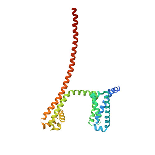
5VB2

Crystal structure of the NavAb voltage-gated sodium channel in a closed conformation


Experimental Data Snapshot

  • Method: X-RAY DIFFRACTION
  • Resolution: 3.20 Å
  • R-Value Free: 0.266 
  • R-Value Work: 0.233 
  • R-Value Observed: 0.235 

Starting Model: experimental
View more details

wwPDB Validation   3D Report Full Report


Ligand Structure Quality Assessment 


This is version 1.3 of the entry. See complete history


Literature

Structures of closed and open states of a voltage-gated sodium channel.

Lenaeus, M.J.Gamal El-Din, T.M.Ing, C.Ramanadane, K.Pomes, R.Zheng, N.Catterall, W.A.

(2017) Proc Natl Acad Sci U S A 114: E3051-E3060

  • DOI: https://doi.org/10.1073/pnas.1700761114
  • Primary Citation of Related Structures:  
    5VB2, 5VB8

  • PubMed Abstract: 

    Bacterial voltage-gated sodium channels (BacNavs) serve as models of their vertebrate counterparts. BacNavs contain conserved voltage-sensing and pore-forming domains, but they are homotetramers of four identical subunits, rather than pseudotetramers of four homologous domains. Here, we present structures of two Na V Ab mutants that capture tightly closed and open states at a resolution of 2.8-3.2 Å. Introduction of two humanizing mutations in the S6 segment (Na V Ab/FY: T206F and V213Y) generates a persistently closed form of the activation gate in which the intracellular ends of the four S6 segments are drawn tightly together to block ion permeation completely. This construct also revealed the complete structure of the four-helix bundle that forms the C-terminal domain. In contrast, truncation of the C-terminal 40 residues in Na v Ab/1-226 captures the activation gate in an open conformation, revealing the open state of a BacNav with intact voltage sensors. Comparing these structures illustrates the full range of motion of the activation gate, from closed with its orifice fully occluded to open with an orifice of ∼10 Å. Molecular dynamics and free-energy simulations confirm designation of Na V Ab/1-226 as an open state that allows permeation of hydrated Na + , and these results also support a hydrophobic gating mechanism for control of ion permeation. These two structures allow completion of a closed-open-inactivated conformational cycle in a single voltage-gated sodium channel and give insight into the structural basis for state-dependent binding of sodium channel-blocking drugs.


  • Organizational Affiliation

    Department of Pharmacology, University of Washington, Seattle, WA 98195.


Macromolecules
Find similar proteins by:  (by identity cutoff)  |  3D Structure
Entity ID: 1
MoleculeChains Sequence LengthOrganismDetailsImage
Ion transport protein
A, B, C, D
285Aliarcobacter butzleriMutation(s): 2 
Membrane Entity: Yes 
UniProt
Find proteins for A8EVM5 (Aliarcobacter butzleri (strain RM4018))
Explore A8EVM5 
Go to UniProtKB:  A8EVM5
Entity Groups  
Sequence Clusters30% Identity50% Identity70% Identity90% Identity95% Identity100% Identity
UniProt GroupA8EVM5
Sequence Annotations
Expand
  • Reference Sequence
Small Molecules
Ligands 4 Unique
IDChains Name / Formula / InChI Key2D Diagram3D Interactions
PX4
Query on PX4

Download Ideal Coordinates CCD File 
AA [auth D]
BA [auth D]
CA [auth D]
G [auth A]
H [auth A]
AA [auth D],
BA [auth D],
CA [auth D],
G [auth A],
H [auth A],
I [auth A],
M [auth B],
N [auth B],
O [auth B],
P [auth B],
T [auth C],
U [auth C],
V [auth C]
1,2-DIMYRISTOYL-SN-GLYCERO-3-PHOSPHOCHOLINE
C36 H73 N O8 P
CITHEXJVPOWHKC-UUWRZZSWSA-O
CPS
Query on CPS

Download Ideal Coordinates CCD File 
E [auth A]
F [auth A]
K [auth B]
L [auth B]
R [auth C]
E [auth A],
F [auth A],
K [auth B],
L [auth B],
R [auth C],
S [auth C],
Y [auth D],
Z [auth D]
3-[(3-CHOLAMIDOPROPYL)DIMETHYLAMMONIO]-1-PROPANESULFONATE
C32 H58 N2 O7 S
UMCMPZBLKLEWAF-BCTGSCMUSA-N
PO4
Query on PO4

Download Ideal Coordinates CCD File 
DA [auth D],
J [auth A],
Q [auth B],
W [auth C]
PHOSPHATE ION
O4 P
NBIIXXVUZAFLBC-UHFFFAOYSA-K
CL
Query on CL

Download Ideal Coordinates CCD File 
X [auth C]CHLORIDE ION
Cl
VEXZGXHMUGYJMC-UHFFFAOYSA-M
Experimental Data & Validation

Experimental Data

  • Method: X-RAY DIFFRACTION
  • Resolution: 3.20 Å
  • R-Value Free: 0.266 
  • R-Value Work: 0.233 
  • R-Value Observed: 0.235 
  • Space Group: P 21 2 21
Unit Cell:
Length ( Å )Angle ( ˚ )
a = 125.963α = 90
b = 129.853β = 90
c = 196.128γ = 90
Software Package:
Software NamePurpose
PHENIXrefinement
HKL-2000data reduction
HKL-2000data scaling
PHASERphasing

Structure Validation

View Full Validation Report


Ligand Structure Quality Assessment 


Entry History 

Deposition Data

Revision History  (Full details and data files)

  • Version 1.0: 2017-04-05
    Type: Initial release
  • Version 1.1: 2017-04-19
    Changes: Database references
  • Version 1.2: 2017-09-27
    Changes: Author supporting evidence
  • Version 1.3: 2023-10-04
    Changes: Data collection, Database references, Refinement description