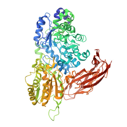
5XXO

Crystal structure of mutant (D286N) GH3 beta-glucosidase from Bacteroides thetaiotaomicron in complex with sophorotriose


Experimental Data Snapshot

  • Method: X-RAY DIFFRACTION
  • Resolution: 2.02 Å
  • R-Value Free: 0.206 
  • R-Value Work: 0.169 
  • R-Value Observed: 0.171 

Starting Model: experimental
View more details

wwPDB Validation   3D Report Full Report


Ligand Structure Quality Assessment 


This is version 2.1 of the entry. See complete history


Literature

Function and structure relationships of a beta-1,2-glucooligosaccharide-degrading beta-glucosidase.

Ishiguro, R.Tanaka, N.Abe, K.Nakajima, M.Maeda, T.Miyanaga, A.Takahashi, Y.Sugimoto, N.Nakai, H.Taguchi, H.

(2017) FEBS Lett 591: 3926-3936

  • DOI: https://doi.org/10.1002/1873-3468.12911
  • Primary Citation of Related Structures:  
    5XXL, 5XXM, 5XXN, 5XXO

  • PubMed Abstract: 

    BT_3567 protein, a putative β-glucosidase from Bacteroides thetaiotaomicron, exhibits higher activity toward Sop 3-5 (Sop n , n: degree of polymerization of β-1,2-glucooligosaccharides) than toward Sop 2 , unlike a known β-glucosidase from Listeria innocua which predominantly prefers Sop 2 . In the complex structure determined by soaking of a D286N mutant crystal with Sop 4 , a Sop 3 moiety was observed at subsites -1 to +2. The glucose moiety at subsite +2 forms a hydrogen bond with Asn81, which is replaced with Gly in the L. innocua β-glucosidase. The K m values of the N81G mutant for Sop 3-5 are much higher than those of the wild-type, suggesting that Asn81 contributes to the binding to substrates longer than Sop 3 .


  • Organizational Affiliation

    Department of Applied Biological Science, Faculty of Science and Technology, Tokyo University of Science, Noda, Chiba, Japan.


Macromolecules
Find similar proteins by:  (by identity cutoff)  |  3D Structure
Entity ID: 1
MoleculeChains Sequence LengthOrganismDetailsImage
Periplasmic beta-glucosidase
A, B
760Bacteroides thetaiotaomicron VPI-5482Mutation(s): 1 
Gene Names: BT_3567
EC: 3.2.1.21
UniProt
Find proteins for Q8A1U1 (Bacteroides thetaiotaomicron (strain ATCC 29148 / DSM 2079 / JCM 5827 / CCUG 10774 / NCTC 10582 / VPI-5482 / E50))
Explore Q8A1U1 
Go to UniProtKB:  Q8A1U1
Entity Groups  
Sequence Clusters30% Identity50% Identity70% Identity90% Identity95% Identity100% Identity
UniProt GroupQ8A1U1
Sequence Annotations
Expand
  • Reference Sequence
Oligosaccharides

Help

Entity ID: 2
MoleculeChains Length2D Diagram Glycosylation3D Interactions
beta-D-glucopyranose-(1-2)-beta-D-glucopyranose-(1-2)-beta-D-glucopyranose
C
3N/A
Glycosylation Resources
GlyTouCan:  G74665DT
GlyCosmos:  G74665DT
GlyGen:  G74665DT
Small Molecules
Ligands 4 Unique
IDChains Name / Formula / InChI Key2D Diagram3D Interactions
PG4
Query on PG4

Download Ideal Coordinates CCD File 
G [auth A]TETRAETHYLENE GLYCOL
C8 H18 O5
UWHCKJMYHZGTIT-UHFFFAOYSA-N
BGC
Query on BGC

Download Ideal Coordinates CCD File 
E [auth A]beta-D-glucopyranose
C6 H12 O6
WQZGKKKJIJFFOK-VFUOTHLCSA-N
PEG
Query on PEG

Download Ideal Coordinates CCD File 
F [auth A]DI(HYDROXYETHYL)ETHER
C4 H10 O3
MTHSVFCYNBDYFN-UHFFFAOYSA-N
MG
Query on MG

Download Ideal Coordinates CCD File 
D [auth A],
H [auth B]
MAGNESIUM ION
Mg
JLVVSXFLKOJNIY-UHFFFAOYSA-N
Experimental Data & Validation

Experimental Data

  • Method: X-RAY DIFFRACTION
  • Resolution: 2.02 Å
  • R-Value Free: 0.206 
  • R-Value Work: 0.169 
  • R-Value Observed: 0.171 
  • Space Group: C 2 2 21
Unit Cell:
Length ( Å )Angle ( ˚ )
a = 82.062α = 90
b = 167.887β = 90
c = 225.45γ = 90
Software Package:
Software NamePurpose
REFMACrefinement
HKL-2000data reduction
MOLREPphasing

Structure Validation

View Full Validation Report


Ligand Structure Quality Assessment 


Entry History 

Deposition Data

Revision History  (Full details and data files)

  • Version 1.0: 2017-12-13
    Type: Initial release
  • Version 1.1: 2017-12-27
    Changes: Database references
  • Version 2.0: 2020-07-29
    Type: Remediation
    Reason: Carbohydrate remediation
    Changes: Atomic model, Data collection, Derived calculations, Structure summary
  • Version 2.1: 2023-11-22
    Changes: Data collection, Database references, Derived calculations, Refinement description, Structure summary