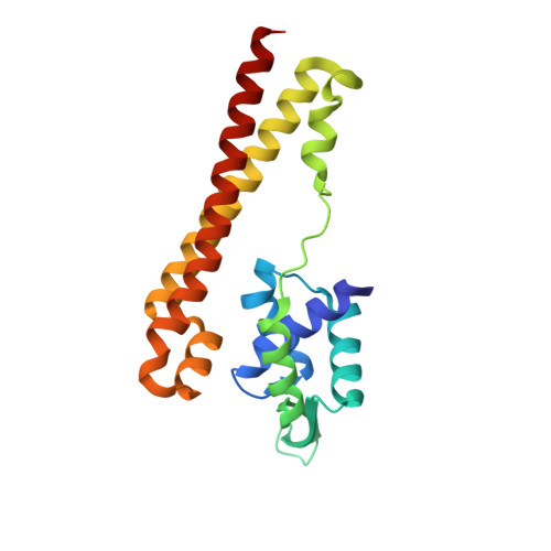
Crystal structure of the VanR transcription factor and the role of its unique alpha-helix in effector recognition.
Kwak, Y.M., Park, S.C., Na, H.W., Kang, S.G., Lee, G.S., Ko, H.J., Kim, P.H., Oh, B.C., Yoon, S.I.(2018) FEBS J 285: 3786-3800
- PubMed: 30095229
- DOI: https://doi.org/10.1111/febs.14629
- Primary Citation of Related Structures:
5Z7B - PubMed Abstract:
VanR is a negative transcriptional regulator of bacteria that belongs to the PadR family and modulates the expression of vanillate transport and degradation proteins in response to vanillate. Although VanR plays a key role in the utilization of vanillate as a carbon source, it is barely understood how VanR recognizes its effector. Thus, our knowledge concerning the gene regulatory mechanism of VanR is limited. Here, we reveal the vanillate-binding mode of VanR through structural, biophysical, and mutational studies. Similar to other PadR family members, VanR forms a functional dimer, and each VanR subunit consists of an N-terminal DNA-binding domain (NTD) and a C-terminal dimerization domain (CTD). One VanR dimer simultaneously binds two vanillate molecules using two interdomain cavities, as observed in PadR. In contrast to these common features, VanR contains an additional α-helix, αi, that has not been found in other PadR family members. The αi helix functions as an interdomain crosslinker that mediates interactions between the NTD and the CTD. In addition, the VanR-specific αi helix plays a key role in the formation of a unique effector-binding site. As a result, the effector-binding mode of VanR is distinguishable from that of PadR in the location and accessibility of the effector-binding site as well as the orientation of its bound effector. Furthermore, we propose the DNA-binding mode and vanillate-mediated transcriptional regulation mechanism of VanR based on comparative structural and mutational analyses. DATABASES: The atomic coordinates and the structure factors for VanR (PDB ID 5Z7B) have been deposited in the Protein Data Bank, www.pdb.org.
Organizational Affiliation:
Division of Biomedical Convergence, College of Biomedical Science, Kangwon National University, Chuncheon, Korea.


