5ZIN

Crystal structure of bacteriorhodopsin at 1.27 A resolution


Experimental Data Snapshot

  • Method: X-RAY DIFFRACTION
  • Resolution: 1.27 Å
  • R-Value Free: 0.163 
  • R-Value Work: 0.130 

Starting Model: experimental
View more details

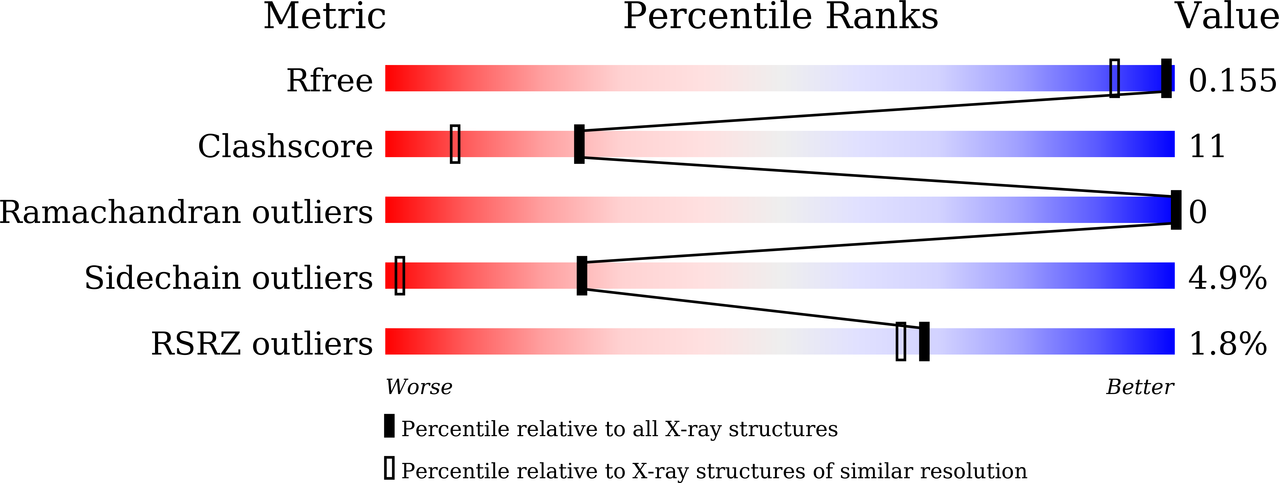
wwPDB Validation   3D Report Full Report


Ligand Structure Quality Assessment 

Created with Raphaël 2.3.0Worse 01 BetterLigand structure goodness of fit to experimental dataBest fitted L2PClick on this verticalbar to view detailsBest fitted RETClick on this verticalbar to view details

This is version 2.2 of the entry. See complete history


Literature

X-ray structure analysis of bacteriorhodopsin at 1.3 angstrom resolution.

Hasegawa, N.Jonotsuka, H.Miki, K.Takeda, K.

(2018) Sci Rep 8: 13123-13123

  • DOI: https://doi.org/10.1038/s41598-018-31370-0
  • Primary Citation of Related Structures:  
    5ZIL, 5ZIM, 5ZIN

  • PubMed Abstract: 

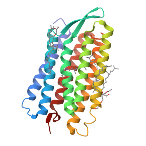
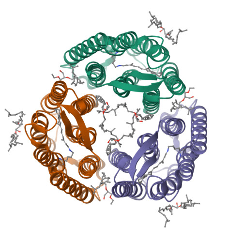
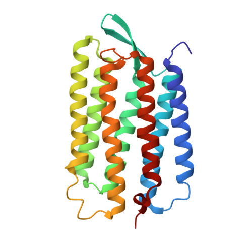
    Bacteriorhodopsin (bR) of Halobacterium salinarum is a membrane protein that acts as a light-driven proton pump. bR and its homologues have recently been utilized in optogenetics and other applications. Although the structures of those have been reported so far, the resolutions are not sufficient for elucidation of the intrinsic structural features critical to the color tuning and ion pumping properties. Here we report the accurate crystallographic analysis of bR in the ground state. The influence of X-rays was suppressed by collecting the data under a low irradiation dose at 15 K. Consequently, individual atoms could be separately observed in the electron density map at better than 1.3 Å resolution. Residues from Thr5 to Ala233 were continuously constructed in the model. The twist of the retinal polyene was determined to be different from those in the previous models. Two conformations were observed for the proton release region. We discuss the meaning of these fine structural features.


  • Organizational Affiliation

    Department of Chemistry, Graduate School of Science, Kyoto University, Sakyo-ku, Kyoto, 606-8502, Japan.


Macromolecules
Find similar proteins by:  (by identity cutoff)  |  3D Structure
Entity ID: 1
MoleculeChains Sequence LengthOrganismDetailsImage
Bacteriorhodopsin228Halobacterium salinarum NRC-1Mutation(s): 0 
Membrane Entity: Yes 
UniProt
Find proteins for P02945 (Halobacterium salinarum (strain ATCC 700922 / JCM 11081 / NRC-1))
Explore P02945 
Go to UniProtKB:  P02945
Entity Groups  
Sequence Clusters30% Identity50% Identity70% Identity90% Identity95% Identity100% Identity
UniProt GroupP02945
Sequence Annotations
Expand
  • Reference Sequence
Experimental Data & Validation

Experimental Data

  • Method: X-RAY DIFFRACTION
  • Resolution: 1.27 Å
  • R-Value Free: 0.163 
  • R-Value Work: 0.130 
  • Space Group: P 63
Unit Cell:
Length ( Å )Angle ( ˚ )
a = 60.63α = 90
b = 60.63β = 90
c = 110.64γ = 120
Software Package:
Software NamePurpose
XSCALEdata scaling
SHELXLrefinement
PDB_EXTRACTdata extraction

Structure Validation

View Full Validation Report


Ligand Structure Quality Assessment 

Created with Raphaël 2.3.0Worse 01 BetterLigand structure goodness of fit to experimental dataBest fitted L2PClick on this verticalbar to view detailsBest fitted RETClick on this verticalbar to view details

Entry History & Funding Information

Deposition Data


Funding OrganizationLocationGrant Number
JSPSJapan25440020, 17H03643

Revision History  (Full details and data files)

  • Version 1.0: 2018-10-10
    Type: Initial release
  • Version 2.0: 2021-05-05
    Changes: Advisory, Atomic model, Author supporting evidence, Data collection, Derived calculations, Other, Structure summary
  • Version 2.1: 2023-11-22
    Changes: Data collection, Database references, Refinement description
  • Version 2.2: 2024-10-30
    Changes: Structure summary