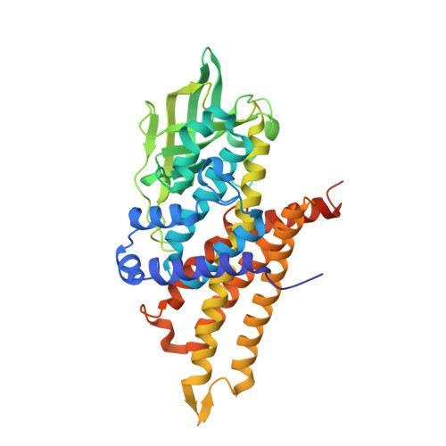
6AF6

PigA with FAD and proline


Experimental Data Snapshot

  • Method: X-RAY DIFFRACTION
  • Resolution: 1.62 Å
  • R-Value Free: 0.180 
  • R-Value Work: 0.155 
  • R-Value Observed: 0.156 

Starting Model: experimental
View more details

wwPDB Validation   3D Report Full Report


Ligand Structure Quality Assessment 


This is version 1.2 of the entry. See complete history


Literature

Crystal Structure of PigA: A Prolyl Thioester-Oxidizing Enzyme in Prodigiosin Biosynthesis.

Lee, C.-C.Ko, T.-P.Chen, C.T.Chan, Y.T.Lo, S.Y.Chang, J.Y.Chen, Y.W.Chung, T.F.Hsieh, H.J.Hsiao, C.D.Wang, A.H.J.

(2019) Chembiochem 20: 193-202

  • DOI: https://doi.org/10.1002/cbic.201800409
  • Primary Citation of Related Structures:  
    5ZW0, 5ZW2, 5ZW7, 5ZW8, 6AF6

  • PubMed Abstract: 

    Prodigiosin is an intensely red pigment comprising three pyrroles. The biosynthetic pathway includes a two-step proline oxidation catalyzed by phosphatidylinositol N-acetylglucosaminyltransferase subunit A (PigA), with flavin adenine dinucleotide (FAD) as its cofactor. The enzyme is crystallized in the apo form and in complex with FAD and proline. As an acyl coenzyme A dehydrogenase (ACAD) family member, the protein folds into a β-sheet flanked by two α-helical domains. PigA forms a tetramer, which is consistent with analytical ultracentrifugation results. FAD binds to PigA in a similar way to that in the other enzymes of the ACAD family. The variable conformations of loop β4-β5 and helix αG correlate well with the structural flexibility required for substrate entrance to the Re side of FAD. Modeling with PigG, the acyl carrier protein, suggests a reasonable mode of interaction with PigA. The structure helps to explain the proline oxidation mechanism, in which Glu244 plays a central role by abstracting the substrate protons. It also reveals a plausible pocket for oxygen binding to the Si side of FAD.


  • Organizational Affiliation

    Institute of Biological Chemistry, Academia Sinica, 128 Academia Road Section 2, Taipei, 11529, Taiwan.


Macromolecules
Find similar proteins by:  (by identity cutoff)  |  3D Structure
Entity ID: 1
MoleculeChains Sequence LengthOrganismDetailsImage
L-prolyl-[peptidyl-carrier protein] dehydrogenase402Prodigiosinella confusarubidaMutation(s): 1 
Gene Names: pigA
EC: 1.3.8.14
UniProt
Find proteins for Q5W271 (Serratia sp. (strain ATCC 39006))
Explore Q5W271 
Go to UniProtKB:  Q5W271
Entity Groups  
Sequence Clusters30% Identity50% Identity70% Identity90% Identity95% Identity100% Identity
UniProt GroupQ5W271
Sequence Annotations
Expand
  • Reference Sequence
Small Molecules
Ligands 5 Unique
IDChains Name / Formula / InChI Key2D Diagram3D Interactions
FAD (Subject of Investigation/LOI)
Query on FAD

Download Ideal Coordinates CCD File 
B [auth A]FLAVIN-ADENINE DINUCLEOTIDE
C27 H33 N9 O15 P2
VWWQXMAJTJZDQX-UYBVJOGSSA-N
O4B
Query on O4B

Download Ideal Coordinates CCD File 
C [auth A],
D [auth A]
1,4,7,10,13,16-HEXAOXACYCLOOCTADECANE
C12 H24 O6
XEZNGIUYQVAUSS-UHFFFAOYSA-N
PRO (Subject of Investigation/LOI)
Query on PRO

Download Ideal Coordinates CCD File 
F [auth A],
G [auth A]
PROLINE
C5 H9 N O2
ONIBWKKTOPOVIA-BYPYZUCNSA-N
GLY (Subject of Investigation/LOI)
Query on GLY

Download Ideal Coordinates CCD File 
H [auth A]GLYCINE
C2 H5 N O2
DHMQDGOQFOQNFH-UHFFFAOYSA-N
CL
Query on CL

Download Ideal Coordinates CCD File 
E [auth A]CHLORIDE ION
Cl
VEXZGXHMUGYJMC-UHFFFAOYSA-M
Experimental Data & Validation

Experimental Data

  • Method: X-RAY DIFFRACTION
  • Resolution: 1.62 Å
  • R-Value Free: 0.180 
  • R-Value Work: 0.155 
  • R-Value Observed: 0.156 
  • Space Group: I 2 2 2
Unit Cell:
Length ( Å )Angle ( ˚ )
a = 85.574α = 90
b = 86.306β = 90
c = 145.771γ = 90
Software Package:
Software NamePurpose
PHENIXrefinement
HKL-2000data reduction
HKL-2000data scaling
PHENIXphasing

Structure Validation

View Full Validation Report


Ligand Structure Quality Assessment 


Entry History & Funding Information

Deposition Data


Funding OrganizationLocationGrant Number
Academia Sinica (Taiwan)Taiwan--

Revision History  (Full details and data files)

  • Version 1.0: 2018-09-05
    Type: Initial release
  • Version 1.1: 2019-02-06
    Changes: Data collection, Database references
  • Version 1.2: 2023-11-22
    Changes: Data collection, Database references, Derived calculations, Refinement description