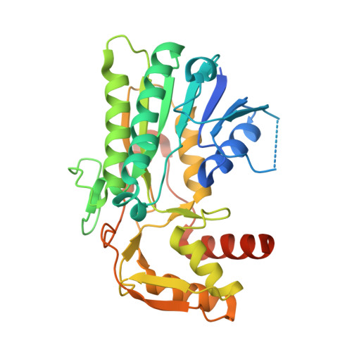
Structure of an antibiotic-synthesizing UDP-glucuronate 4-epimerase MoeE5 in complex with substrate.
Sun, H., Ko, T.P., Liu, W., Liu, W., Zheng, Y., Chen, C.C., Guo, R.T.(2020) Biochem Biophys Res Commun 521: 31-36
- PubMed: 31653344
- DOI: https://doi.org/10.1016/j.bbrc.2019.10.035
- Primary Citation of Related Structures:
6KV9, 6KVC - PubMed Abstract:
The epimerase MoeE5 from Streptomyces viridosporus converts UDP-glucuronic acid (UDP-GlcA) to UDP-galacturonic acid (UDP-GalA) to provide the first sugar in synthesizing moenomycin, a potent inhibitor against bacterial peptidoglycan glycosyltransferases. The enzyme belongs to the UDP-hexose 4-epimerase family, and uses NAD + as its cofactor. Here we present the complex crystal structures of MoeE5/NAD + /UDP-GlcA and MoeE5/NAD + /UDP-glucose, determined at 1.48 Å and 1.66 Å resolution. The cofactor NAD + is bound to the N-terminal Rossmann-fold domain and the substrate is bound to the smaller C-terminal domain. In both crystals the C4 atom of the sugar moiety of the substrate is in close proximity to the C4 atom of the nicotinamide of NAD + , and the O4 atom of the sugar is also hydrogen bonded to the side chain of Tyr154, suggesting a productive binding mode. As the first complex structure of this protein family with a bound UDP-GlcA in the active site, it shows an extensive hydrogen-bond network between the enzyme and the substrate. We further built a model with the product UDP-GalA, and found that the unique Arg192 of MoeE5 might play an important role in the catalytic pathway. Consequently, MoeE5 is likely a specific epimerase for UDP-GlcA to UDP-GalA conversion, rather than a promiscuous enzyme as some other family members.
Organizational Affiliation:
University of Chinese Academy of Sciences, Beijing, 100049, China; Tianjin Institute of Industrial Biotechnology, Chinese Academy of Sciences, Tianjin, 300308, China.


