6MLW | pdb_00006mlw

Crystal structure of X. citri phosphoglucomutase in complex with 2-fluoro mannosyl-1-methyl-phosphonic acid


Experimental Data Snapshot

  • Method: X-RAY DIFFRACTION
  • Resolution: 1.90 Å
  • R-Value Free: 
    0.249 (Depositor), 0.250 (DCC) 
  • R-Value Work: 
    0.189 (Depositor), 0.190 (DCC) 
  • R-Value Observed: 
    0.192 (Depositor) 

Starting Model: experimental
View more details

wwPDB Validation   3D Report Full Report


Ligand Structure Quality Assessment 

Created with Raphaël 2.3.0Worse 01 BetterLigand structure goodness of fit to experimental dataBest fitted JVAClick on this verticalbar to view details

This is version 1.5 of the entry. See complete history


Literature

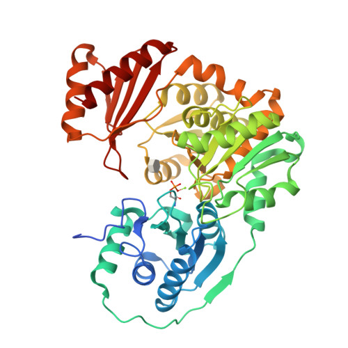
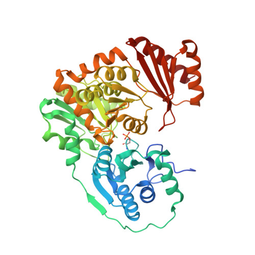
Inhibitory Evaluation of alpha PMM/PGM fromPseudomonas aeruginosa: Chemical Synthesis, Enzyme Kinetics, and Protein Crystallographic Study.

Zhu, J.S.Stiers, K.M.Soleimani, E.Groves, B.R.Beamer, L.J.Jakeman, D.L.

(2019) J Org Chem 84: 9627-9636

  • DOI: https://doi.org/10.1021/acs.joc.9b01305
  • Primary Citation of Related Structures:  
    6MLF, 6MLH, 6MLW, 6MNV

  • PubMed Abstract: 

    α-Phosphomannomutase/phosphoglucomutase (αPMM/PGM) from P. aeruginosa is involved in bacterial cell wall assembly and is implicated in P. aeruginosa virulence, yet few studies have addressed αPMM/PGM inhibition from this important Gram-negative bacterial human pathogen. Four structurally different α-d-glucopyranose 1-phosphate (αG1P) derivatives including 1- C -fluoromethylated analogues ( 1 - 3 ), 1,2-cyclic phosph(on)ate analogues ( 4 - 6 ), isosteric methylene phosphono analogues ( 7 and 8 ), and 6-fluoro-αG1P ( 9 ), were synthesized and assessed as potential time-dependent or reversible αPMM/PGM inhibitors. The resulting kinetic data were consistent with the crystallographic structures of the highly homologous Xanthomonas citri αPGM with inhibitors 3 and 7 - 9 binding to the enzyme active site (1.65-1.9 Å). These structural and kinetic insights will enhance the design of future αPMM/PGM inhibitors.


  • Organizational Affiliation

    College of Pharmacy , Dalhousie University , 5968 College Street , Halifax , Nova Scotia B3H 4R2 , Canada.


Macromolecules
Find similar proteins by:  (by identity cutoff)  |  3D Structure
Entity ID: 1
MoleculeChains Sequence LengthOrganismDetailsImage
Phosphoglucomutase468Xanthomonas citriMutation(s): 0 
Gene Names: xanA
Entity Groups  
Sequence Clusters30% Identity50% Identity70% Identity90% Identity95% Identity100% Identity
Sequence Annotations
Expand
  • Reference Sequence
Small Molecules
Ligands 2 Unique
IDChains Name / Formula / InChI Key2D Diagram3D Interactions
JVA
Query on JVA

Download Ideal Coordinates CCD File 
C [auth A]2,6-anhydro-5,7-dideoxy-5-fluoro-7-phosphono-D-glycero-D-manno-heptitol
C7 H14 F O7 P
CDQMFQZEPGCBTC-NYMZXIIRSA-N
MG
Query on MG

Download Ideal Coordinates CCD File 
B [auth A]MAGNESIUM ION
Mg
JLVVSXFLKOJNIY-UHFFFAOYSA-N
Modified Residues  1 Unique
IDChains TypeFormula2D DiagramParent
SEP
Query on SEP
A
L-PEPTIDE LINKINGC3 H8 N O6 PSER
Experimental Data & Validation

Experimental Data

  • Method: X-RAY DIFFRACTION
  • Resolution: 1.90 Å
  • R-Value Free:  0.249 (Depositor), 0.250 (DCC) 
  • R-Value Work:  0.189 (Depositor), 0.190 (DCC) 
  • R-Value Observed: 0.192 (Depositor) 
Space Group: P 21 21 21
Unit Cell:
Length ( Å )Angle ( ˚ )
a = 43.781α = 90
b = 54.587β = 90
c = 172.909γ = 90
Software Package:
Software NamePurpose
PHENIXrefinement
XDSdata reduction
SCALAdata scaling
PHENIXphasing

Structure Validation

View Full Validation Report


Ligand Structure Quality Assessment 

Created with Raphaël 2.3.0Worse 01 BetterLigand structure goodness of fit to experimental dataBest fitted JVAClick on this verticalbar to view details

Entry History & Funding Information

Deposition Data


Funding OrganizationLocationGrant Number
National Science Foundation (NSF, United States)United StatesMCB 1409898

Revision History  (Full details and data files)

  • Version 1.0: 2019-07-31
    Type: Initial release
  • Version 1.1: 2019-08-14
    Changes: Data collection, Database references
  • Version 1.2: 2019-11-27
    Changes: Author supporting evidence
  • Version 1.3: 2020-07-29
    Type: Remediation
    Reason: Carbohydrate remediation
    Changes: Data collection, Derived calculations
  • Version 1.4: 2023-10-11
    Changes: Data collection, Database references, Refinement description
  • Version 1.5: 2024-10-23
    Changes: Structure summary