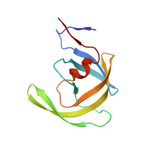
6OPS

HIV-1 Protease NL4-3 WT in complex with darunavir


Experimental Data Snapshot

  • Method: X-RAY DIFFRACTION
  • Resolution: 2.08 Å
  • R-Value Free: 0.251 
  • R-Value Work: 0.225 
  • R-Value Observed: 0.225 

Starting Model: experimental
View more details

wwPDB Validation   3D Report Full Report


Ligand Structure Quality Assessment 


This is version 1.3 of the entry. See complete history


Literature

Picomolar to Micromolar: Elucidating the Role of Distal Mutations in HIV-1 Protease in Conferring Drug Resistance.

Henes, M.Lockbaum, G.J.Kosovrasti, K.Leidner, F.Nachum, G.S.Nalivaika, E.A.Lee, S.K.Spielvogel, E.Zhou, S.Swanstrom, R.Bolon, D.N.A.Kurt Yilmaz, N.Schiffer, C.A.

(2019) ACS Chem Biol 14: 2441-2452

  • DOI: https://doi.org/10.1021/acschembio.9b00370
  • Primary Citation of Related Structures:  
    6OPS, 6OPT, 6OPU, 6OPV, 6OPW, 6OPX, 6OPY, 6OPZ

  • PubMed Abstract: 

    Drug resistance continues to be a growing global problem. The efficacy of small molecule inhibitors is threatened by pools of genetic diversity in all systems, including antibacterials, antifungals, cancer therapeutics, and antivirals. Resistant variants often include combinations of active site mutations and distal "secondary" mutations, which are thought to compensate for losses in enzymatic activity. HIV-1 protease is the ideal model system to investigate these combinations and underlying molecular mechanisms of resistance. Darunavir (DRV) binds wild-type (WT) HIV-1 protease with a potency of <5 pM, but we have identified a protease variant that loses potency to DRV 150 000-fold, with 11 mutations in and outside the active site. To elucidate the roles of these mutations in DRV resistance, we used a multidisciplinary approach, combining enzymatic assays, crystallography, and molecular dynamics simulations. Analysis of protease variants with 1, 2, 4, 8, 9, 10, and 11 mutations showed that the primary active site mutations caused ∼50-fold loss in potency (2 mutations), while distal mutations outside the active site further decreased DRV potency from 13 nM (8 mutations) to 0.76 μM (11 mutations). Crystal structures and simulations revealed that distal mutations induce subtle changes that are dynamically propagated through the protease. Our results reveal that changes remote from the active site directly and dramatically impact the potency of the inhibitor. Moreover, we find interdependent effects of mutations in conferring high levels of resistance. These mechanisms of resistance are likely applicable to many other quickly evolving drug targets, and the insights may have implications for the design of more robust inhibitors.


  • Organizational Affiliation

    Department of Biochemistry and Molecular Pharmacology , University of Massachusetts Medical School , Worcester , Massachusetts 01605 , United States.


Macromolecules
Find similar proteins by:  (by identity cutoff)  |  3D Structure
Entity ID: 1
MoleculeChains Sequence LengthOrganismDetailsImage
Protease NL4-3A [auth B],
B [auth A]
99Human immunodeficiency virus 1Mutation(s): 1 
Gene Names: pol
UniProt
Find proteins for Q8ULI9 (Human immunodeficiency virus type 1)
Explore Q8ULI9 
Go to UniProtKB:  Q8ULI9
Entity Groups  
Sequence Clusters30% Identity50% Identity70% Identity90% Identity95% Identity100% Identity
UniProt GroupQ8ULI9
Sequence Annotations
Expand
  • Reference Sequence
Small Molecules
Ligands 1 Unique
IDChains Name / Formula / InChI Key2D Diagram3D Interactions
017 (Subject of Investigation/LOI)
Query on 017

Download Ideal Coordinates CCD File 
C [auth A](3R,3AS,6AR)-HEXAHYDROFURO[2,3-B]FURAN-3-YL(1S,2R)-3-[[(4-AMINOPHENYL)SULFONYL](ISOBUTYL)AMINO]-1-BENZYL-2-HYDROXYPROPYLCARBAMATE
C27 H37 N3 O7 S
CJBJHOAVZSMMDJ-HEXNFIEUSA-N
Binding Affinity Annotations 
IDSourceBinding Affinity
017 BindingDB:  6OPS Ki: min: 1.47e-4, max: 1.3 (nM) from 27 assay(s)
Kd: 0.02 (nM) from 1 assay(s)
IC50: min: 0.01, max: 4 (nM) from 14 assay(s)
EC50: min: 13, max: 112 (nM) from 2 assay(s)
Experimental Data & Validation

Experimental Data

  • Method: X-RAY DIFFRACTION
  • Resolution: 2.08 Å
  • R-Value Free: 0.251 
  • R-Value Work: 0.225 
  • R-Value Observed: 0.225 
  • Space Group: P 61
Unit Cell:
Length ( Å )Angle ( ˚ )
a = 62.276α = 90
b = 62.276β = 90
c = 82.266γ = 120
Software Package:
Software NamePurpose
HKL-3000data scaling
PHASERphasing
PHENIXrefinement
Cootmodel building

Structure Validation

View Full Validation Report


Ligand Structure Quality Assessment 


Entry History & Funding Information

Deposition Data


Funding OrganizationLocationGrant Number
National Institutes of Health/National Institute of General Medical Sciences (NIH/NIGMS)United StatesP01-GM109767

Revision History  (Full details and data files)

  • Version 1.0: 2019-09-04
    Type: Initial release
  • Version 1.1: 2019-11-27
    Changes: Database references
  • Version 1.2: 2020-01-01
    Changes: Author supporting evidence
  • Version 1.3: 2023-10-11
    Changes: Data collection, Database references, Refinement description, Structure summary