6S99

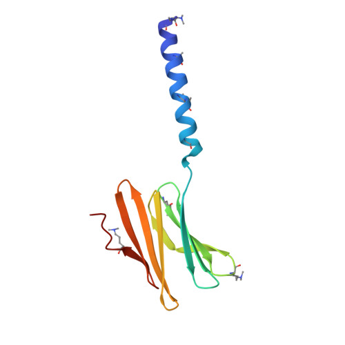
Dimethylated fusion protein of RSL and trimeric coiled coil in complex with cucurbit[7]uril


Experimental Data Snapshot

  • Method: X-RAY DIFFRACTION
  • Resolution: 2.65 Å
  • R-Value Free: 0.257 
  • R-Value Work: 0.225 
  • R-Value Observed: 0.226 

wwPDB Validation   3D Report Full Report


Ligand Structure Quality Assessment 


This is version 2.0 of the entry. See complete history


Literature

Segregated Protein-Cucurbit[7]uril Crystalline Architectures via Modulatory Peptide Tectons.

Ramberg, K.O.Guagnini, F.Engilberge, S.Wronska, M.A.Rennie, M.L.Perez, J.Crowley, P.B.

(2021) Chemistry 

  • DOI: https://doi.org/10.1002/chem.202103025
  • Primary Citation of Related Structures:  
    6S99, 7P2H, 7P2I, 7P2J

  • PubMed Abstract: 

    One approach to protein assembly involves water-soluble supramolecular receptors that act like glues. Bionanoarchitectures directed by these scaffolds are often system-specific, with few studies investigating their customization. Herein, the modulation of cucurbituril-mediated protein assemblies through the inclusion of peptide tectons is described. Three peptides of varying length and structural order were N-terminally appended to RSL, a β-propeller building block. Each fusion protein was incorporated into crystalline architectures mediated by cucurbit[7]uril (Q7). A trimeric coiled-coil served as a spacer within a Q7-directed sheet assembly of RSL, giving rise to a layered material of varying porosity. Within the spacer layers, the coiled-coils were dynamic. This result prompted consideration of intrinsically disordered peptides (IDPs) as modulatory tectons. Similar to the coiled-coil, a mussel adhesion peptide (Mefp) also acted as a spacer between protein-Q7 sheets. In contrast, the fusion of a nucleoporin peptide (Nup) to RSL did not recapitulate the sheet assembly. Instead, a Q7-directed cage was adopted, within which disordered Nup peptides were partially "captured" by Q7 receptors. IDP capture occurred by macrocycle recognition of an intrapeptide Phe-Gly motif in which the benzyl group was encapsulated by Q7. The modularity of these protein-cucurbituril architectures adds a new dimension to macrocycle-mediated protein assembly. Segregated protein crystals, with alternating layers of high and low porosity, could provide a basis for new types of materials.


  • Organizational Affiliation

    School of Chemistry, National University of Ireland Galway, University Road, Galway, H91 TK33, Ireland.


Macromolecules
Find similar proteins by:  (by identity cutoff)  |  3D Structure
Entity ID: 1
MoleculeChains Sequence LengthOrganismDetailsImage
4dzn-RSL,Fucose-binding lectin protein,Fucose-binding lectin protein
A, B, C, D, E
A, B, C, D, E, F
114Ralstonia solanacearumMutation(s): 0 
UniProt
Find proteins for A0A0S4TLR1 (Ralstonia solanacearum)
Explore A0A0S4TLR1 
Go to UniProtKB:  A0A0S4TLR1
Entity Groups  
Sequence Clusters30% Identity50% Identity70% Identity90% Identity95% Identity100% Identity
UniProt GroupA0A0S4TLR1
Sequence Annotations
Expand
  • Reference Sequence
Small Molecules
Ligands 2 Unique
IDChains Name / Formula / InChI Key2D Diagram3D Interactions
QQ7 (Subject of Investigation/LOI)
Query on QQ7

Download Ideal Coordinates CCD File 
I [auth A]
L [auth B]
O [auth C]
R [auth D]
U [auth E]
I [auth A],
L [auth B],
O [auth C],
R [auth D],
U [auth E],
X [auth F]
cucurbit[7]uril
C42 H42 N28 O14
ZDOBFUIMGBWEAB-XGFHMVPTSA-N
BMA
Query on BMA

Download Ideal Coordinates CCD File 
G [auth A]
H [auth A]
J [auth B]
K [auth B]
M [auth C]
G [auth A],
H [auth A],
J [auth B],
K [auth B],
M [auth C],
N [auth C],
P [auth D],
Q [auth D],
S [auth E],
T [auth E],
V [auth F],
W [auth F]
beta-D-mannopyranose
C6 H12 O6
WQZGKKKJIJFFOK-RWOPYEJCSA-N
Modified Residues  1 Unique
IDChains TypeFormula2D DiagramParent
MLY
Query on MLY
A, B, C, D, E
A, B, C, D, E, F
L-PEPTIDE LINKINGC8 H18 N2 O2LYS
Experimental Data & Validation

Experimental Data

  • Method: X-RAY DIFFRACTION
  • Resolution: 2.65 Å
  • R-Value Free: 0.257 
  • R-Value Work: 0.225 
  • R-Value Observed: 0.226 
  • Space Group: P 1
Unit Cell:
Length ( Å )Angle ( ˚ )
a = 51.219α = 76.8
b = 51.242β = 89.88
c = 111.282γ = 60.05
Software Package:
Software NamePurpose
PHENIXrefinement
XDSdata reduction
Aimlessdata scaling
PHASERphasing
PDB_EXTRACTdata extraction

Structure Validation

View Full Validation Report


Ligand Structure Quality Assessment 


Entry History & Funding Information

Deposition Data


Funding OrganizationLocationGrant Number
Science Foundation IrelandIreland--

Revision History  (Full details and data files)

  • Version 1.0: 2020-08-26
    Type: Initial release
  • Version 1.1: 2021-09-08
    Changes: Database references
  • Version 2.0: 2022-12-07
    Changes: Advisory, Atomic model, Data collection, Database references, Derived calculations, Polymer sequence, Source and taxonomy, Structure summary