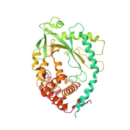
7BUM

mcGAS bound with pGpA


Experimental Data Snapshot

  • Method: X-RAY DIFFRACTION
  • Resolution: 3.05 Å
  • R-Value Free: 0.280 
  • R-Value Work: 0.217 
  • R-Value Observed: 0.223 

Starting Model: experimental
View more details

wwPDB Validation   3D Report Full Report


Ligand Structure Quality Assessment 


This is version 2.0 of the entry. See complete history


Literature

Mn2+Directly Activates cGAS and Structural Analysis Suggests Mn2+Induces a Noncanonical Catalytic Synthesis of 2'3'-cGAMP.

Zhao, Z.Ma, Z.Wang, B.Guan, Y.Su, X.D.Jiang, Z.

(2020) Cell Rep 32: 108053-108053

  • DOI: https://doi.org/10.1016/j.celrep.2020.108053
  • Primary Citation of Related Structures:  
    7BUJ, 7BUM, 7BUQ

  • PubMed Abstract: 

    DNA binding allosterically activates the cytosolic DNA sensor cGAS (cyclic GMP-AMP [cGAMP] synthase) to synthesize 2'3'-cGAMP, using Mg 2+ as the metal cofactor that catalyzes two nucleotidyl-transferring reactions. We previously found that Mn 2+ potentiates cGAS activation, but the underlying mechanism remains unclear. Here, we report that Mn 2+ directly activates cGAS. Structural analysis reveals that Mn 2+ -activated cGAS undergoes globally similar conformational changes to DNA-activated cGAS but forms a unique η1 helix to widen the catalytic pocket, allowing substrate entry and cGAMP synthesis. Strikingly, in Mn 2+ -activated cGAS, the linear intermediates pppGpG and pGpA take an inverted orientation in the active pocket, suggesting a noncanonical but accelerated cGAMP cyclization without substrate flip-over. Moreover, unlike the octahedral coordination around Mg 2+ , the two catalytic Mn 2+ are coordinated by triphosphate moiety of the inverted substrate, independent of the catalytic triad residues. Our findings thus uncover Mn 2+ as a cGAS activator that initiates noncanonical 2'3'-cGAMP synthesis.


  • Organizational Affiliation

    Key Laboratory of Cell Proliferation and Differentiation of the Ministry of Education, School of Life Sciences, Peking University, Beijing 100871, China; Peking-Tsinghua Center for Life Sciences, Peking University, Beijing 100871, China.


Macromolecules
Find similar proteins by:  (by identity cutoff)  |  3D Structure
Entity ID: 1
MoleculeChains Sequence LengthOrganismDetailsImage
Cyclic GMP-AMP synthase
A, B
507Mus musculusMutation(s): 0 
Gene Names: CgasMb21d1
EC: 2.7.7.86
UniProt & NIH Common Fund Data Resources
Find proteins for Q8C6L5 (Mus musculus)
Explore Q8C6L5 
Go to UniProtKB:  Q8C6L5
IMPC:  MGI:2442261
Entity Groups  
Sequence Clusters30% Identity50% Identity70% Identity90% Identity95% Identity100% Identity
UniProt GroupQ8C6L5
Sequence Annotations
Expand
  • Reference Sequence
Small Molecules
Ligands 3 Unique
IDChains Name / Formula / InChI Key2D Diagram3D Interactions
5GP
Query on 5GP

Download Ideal Coordinates CCD File 
E [auth A],
H [auth B]
GUANOSINE-5'-MONOPHOSPHATE
C10 H14 N5 O8 P
RQFCJASXJCIDSX-UUOKFMHZSA-N
AMP (Subject of Investigation/LOI)
Query on AMP

Download Ideal Coordinates CCD File 
D [auth A],
G [auth B]
ADENOSINE MONOPHOSPHATE
C10 H14 N5 O7 P
UDMBCSSLTHHNCD-KQYNXXCUSA-N
ZN (Subject of Investigation/LOI)
Query on ZN

Download Ideal Coordinates CCD File 
C [auth A],
F [auth B]
ZINC ION
Zn
PTFCDOFLOPIGGS-UHFFFAOYSA-N
Experimental Data & Validation

Experimental Data

  • Method: X-RAY DIFFRACTION
  • Resolution: 3.05 Å
  • R-Value Free: 0.280 
  • R-Value Work: 0.217 
  • R-Value Observed: 0.223 
  • Space Group: P 1 21 1
Unit Cell:
Length ( Å )Angle ( ˚ )
a = 48.337α = 90
b = 109.592β = 93.93
c = 75.87γ = 90
Software Package:
Software NamePurpose
PHENIXrefinement
HKL-2000data reduction
SCALEPACKdata scaling
PHASERphasing
PDB_EXTRACTdata extraction

Structure Validation

View Full Validation Report


Ligand Structure Quality Assessment 


Entry History & Funding Information

Deposition Data


Funding OrganizationLocationGrant Number
National Science Foundation (NSF, China)China31670740
National Science Foundation (NSF, China)China31270803

Revision History  (Full details and data files)

  • Version 1.0: 2020-09-02
    Type: Initial release
  • Version 1.1: 2023-11-29
    Changes: Data collection, Database references, Refinement description
  • Version 2.0: 2024-05-29
    Changes: Advisory, Atomic model, Author supporting evidence, Data collection, Derived calculations, Non-polymer description, Structure summary