7LNE

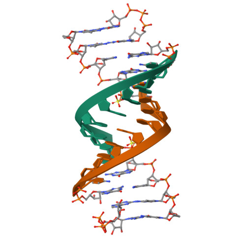
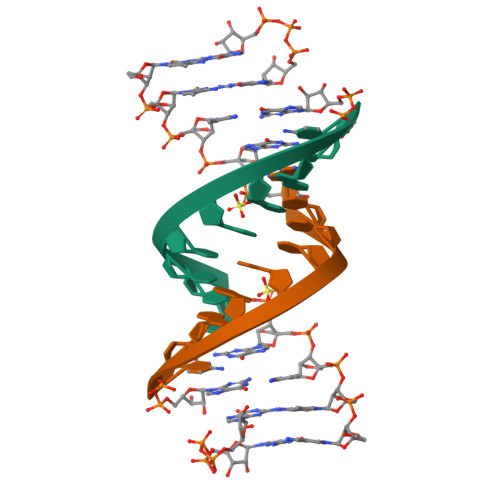
ANA modification at 3' end of RNA primer complex with guanosine dinucleotide ligand G(5')ppp(5')G

  • Classification: RNA
  • Organism(s): synthetic construct
  • Mutation(s): No 

  • Deposited: 2021-02-07 Released: 2021-09-08 
  • Deposition Author(s): Fang, Z., Giurgiu, C., Szostak, J.W.
  • Funding Organization(s): National Science Foundation (NSF, United States), Howard Hughes Medical Institute (HHMI)

Experimental Data Snapshot

  • Method: X-RAY DIFFRACTION
  • Resolution: 1.53 Å
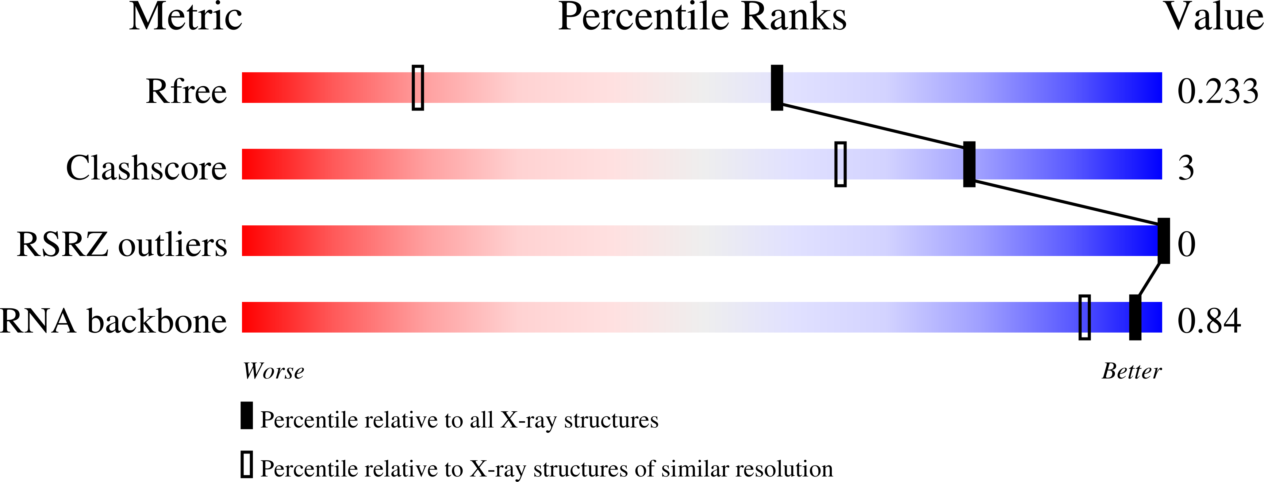
  • R-Value Free: 0.225 
  • R-Value Work: 0.191 

Starting Model: experimental
View more details

wwPDB Validation   3D Report Full Report


Ligand Structure Quality Assessment 


This is version 1.3 of the entry. See complete history


Literature

Structure-Activity Relationships in Nonenzymatic Template-Directed RNA Synthesis.

Giurgiu, C.Fang, Z.Aitken, H.R.M.Kim, S.C.Pazienza, L.Mittal, S.Szostak, J.W.

(2021) Angew Chem Int Ed Engl 60: 22925-22932

  • DOI: https://doi.org/10.1002/anie.202109714
  • Primary Citation of Related Structures:  
    7KUK, 7KUL, 7KUM, 7KUN, 7KUO, 7KUP, 7LNE, 7LNF, 7LNG

  • PubMed Abstract: 

    The template-directed synthesis of RNA played an important role in the transition from prebiotic chemistry to the beginnings of RNA based life, but the mechanism of RNA copying chemistry is incompletely understood. We measured the kinetics of template copying with a set of primers with modified 3'-nucleotides and determined the crystal structures of these modified nucleotides in the context of a primer/template/substrate-analog complex. pH-rate profiles and solvent isotope effects show that deprotonation of the primer 3'-hydroxyl occurs prior to the rate limiting step, the attack of the alkoxide on the activated phosphate of the incoming nucleotide. The analogs with a 3 E ribose conformation show the fastest formation of 3'-5' phosphodiester bonds. Among those derivatives, the reaction rate is strongly correlated with the electronegativity of the 2'-substituent. We interpret our results in terms of differences in steric bulk and charge distribution in the ground vs. transition states.


  • Organizational Affiliation

    Howard Hughes Medical Institute, Department of Molecular Biology, and Center for Computational and Integrative Biology, Massachusetts General Hospital, Boston, MA, 02114, USA.


Macromolecules

Find similar nucleic acids by:  Sequence   |   3D Structure  

Entity ID: 1
MoleculeChains LengthOrganismImage
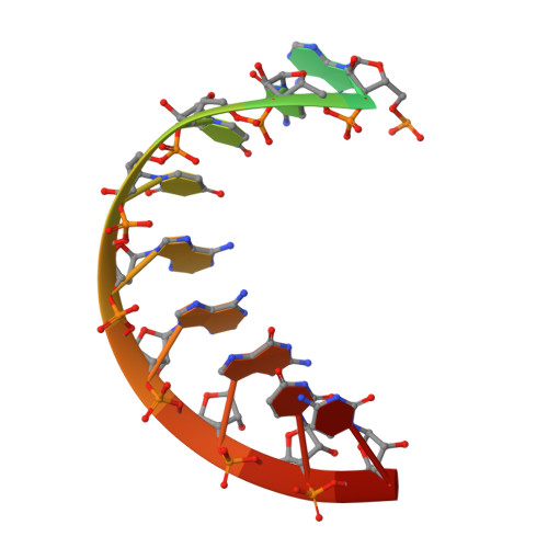
RNA (5'-R(*(LCC)P*(LCC)P*(LCC)P*(LCG)P*AP*CP*UP*UP*AP*AP*GP*UP*CP*(GAO))-3')
A, B
14synthetic construct
Sequence Annotations
Expand
  • Reference Sequence
Small Molecules
Ligands 6 Unique
IDChains Name / Formula / InChI Key2D Diagram3D Interactions
GP3 (Subject of Investigation/LOI)
Query on GP3

Download Ideal Coordinates CCD File 
F [auth A],
O [auth B]
DIGUANOSINE-5'-TRIPHOSPHATE
C20 H27 N10 O18 P3
AAXYAFFKOSNMEB-MHARETSRSA-N
LCG
Query on LCG

Download Ideal Coordinates CCD File 
D [auth A],
M [auth B]
[(1R,3R,4R,7S)-7-HYDROXY-3-(GUANIN-9-YL)-2,5-DIOXABICYCLO[2.2.1]HEPT-1-YL]METHYL DIHYDROGEN PHOSPHATE
C11 H14 N5 O8 P
FZGXOAHULCKQGE-HLJYALQUSA-N
GAO (Subject of Investigation/LOI)
Query on GAO

Download Ideal Coordinates CCD File 
E [auth A],
N [auth B]
GUANINE ARABINOSE-5'-PHOSPHATE
C10 H14 N5 O8 P
RQFCJASXJCIDSX-FJFJXFQQSA-N
LCC
Query on LCC

Download Ideal Coordinates CCD File 
C [auth A],
H [auth A],
K [auth B],
L [auth B]
[(1R,3R,4R,7S)-7-HYDROXY-3-(5-METHYLCYTOSIN-1-YL)-2,5-DIOXABICYCLO[2.2.1]HEPT-1-YL]METHYL DIHYDROGEN PHOSPHATE
C11 H16 N3 O8 P
RSSHBVFUGGVGMZ-YRCORFKGSA-N
LKC
Query on LKC

Download Ideal Coordinates CCD File 
G [auth A],
J [auth B]
4-AMINO-1-[(1S,3R,4R,7S)-7-HYDROXY-1-(HYDROXYMETHYL)-2,5-DIOXABICYCLO[2.2.1]HEPT-3-YL]-5-METHYLPYRIMIDIN-2(1H)-ONE
C11 H15 N3 O5
NELYIRACPIJATH-SZVQBCOZSA-N
SO4
Query on SO4

Download Ideal Coordinates CCD File 
I [auth A],
P [auth B]
SULFATE ION
O4 S
QAOWNCQODCNURD-UHFFFAOYSA-L
Experimental Data & Validation

Experimental Data

  • Method: X-RAY DIFFRACTION
  • Resolution: 1.53 Å
  • R-Value Free: 0.225 
  • R-Value Work: 0.191 
  • Space Group: P 3 2 1
Unit Cell:
Length ( Å )Angle ( ˚ )
a = 43.408α = 90
b = 43.408β = 90
c = 86.554γ = 120
Software Package:
Software NamePurpose
REFMACrefinement
HKL-2000data reduction
HKL-2000data scaling
PHASERphasing

Structure Validation

View Full Validation Report


Ligand Structure Quality Assessment 


Entry History & Funding Information

Deposition Data


Funding OrganizationLocationGrant Number
National Science Foundation (NSF, United States)United States1607034
Howard Hughes Medical Institute (HHMI)United States--

Revision History  (Full details and data files)

  • Version 1.0: 2021-09-08
    Type: Initial release
  • Version 1.1: 2021-10-20
    Changes: Database references
  • Version 1.2: 2022-03-23
    Changes: Database references
  • Version 1.3: 2023-10-18
    Changes: Data collection, Refinement description