7ZS1

Diheme cytochrome c Kustd1711 from Kuenenia stuttgartiensis, M292C mutant


Experimental Data Snapshot

  • Method: X-RAY DIFFRACTION
  • Resolution: 1.70 Å
  • R-Value Free: 
    0.225 (Depositor), 0.220 (DCC) 
  • R-Value Work: 
    0.197 (Depositor), 0.200 (DCC) 
  • R-Value Observed: 
    0.199 (Depositor) 

Starting Model: other
View more details

wwPDB Validation   3D Report Full Report


Ligand Structure Quality Assessment 

Created with Raphaël 2.3.0Worse 01 BetterLigand structure goodness of fit to experimental dataBest fitted HECClick on this verticalbar to view details

This is version 1.4 of the entry. See complete history


Literature

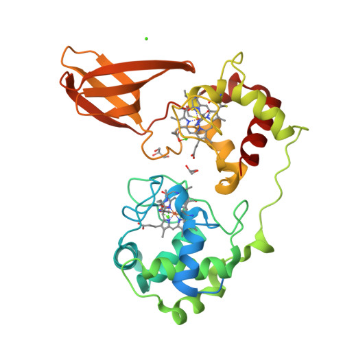
Redox potential tuning by calcium ions in a novel c-type cytochrome from an anammox organism.

Akram, M.Hauser, D.Dietl, A.Steigleder, M.Ullmann, G.M.Barends, T.R.M.

(2024) J Biological Chem : 108082-108082

  • DOI: https://doi.org/10.1016/j.jbc.2024.108082
  • Primary Citation of Related Structures:  
    7ZS0, 7ZS1, 7ZS2, 9FBK

  • PubMed Abstract: 

    The electrochemical potentials of redox-active proteins need to be tuned accurately to the correct values for proper biological function. Here, we describe a diheme cytochrome c with high heme redox potentials of about +350 mV, despite having a large overall negative charge, which typically reduces redox potentials. High-resolution crystal structures, spectroelectrochemical measurements, and high-end computational methods show how this is achieved: each heme iron has a calcium cation positioned next to it at a distance of only 6.9 Å, raising their redox potentials by several hundred millivolts through electrostatic interaction. We suggest that this has evolved to provide the protein with a high redox potential despite its large negative surface charge, which it likely requires for interactions with redox partners.


  • Organizational Affiliation

    Department of Biomolecular Mechanisms, Max Planck Institute for Medical Research, Heidelberg, Germany.


Macromolecules
Find similar proteins by:  (by identity cutoff)  |  3D Structure
Entity ID: 1
MoleculeChains Sequence LengthOrganismDetailsImage
Hypothetical (Diheme) protein316Candidatus KueneniaMutation(s): 1 
Gene Names: KsCSTR_07890KSMBR1_1466kustd1711
UniProt
Find proteins for Q1PZE6 (Kuenenia stuttgartiensis)
Explore Q1PZE6 
Go to UniProtKB:  Q1PZE6
Entity Groups  
Sequence Clusters30% Identity50% Identity70% Identity90% Identity95% Identity100% Identity
UniProt GroupQ1PZE6
Sequence Annotations
Expand
  • Reference Sequence
Small Molecules
Ligands 3 Unique
IDChains Name / Formula / InChI Key2D Diagram3D Interactions
HEC (Subject of Investigation/LOI)
Query on HEC

Download Ideal Coordinates CCD File 
E [auth A],
F [auth A]
HEME C
C34 H34 Fe N4 O4
HXQIYSLZKNYNMH-LJNAALQVSA-N
GOL
Query on GOL

Download Ideal Coordinates CCD File 
G [auth A],
H [auth A]
GLYCEROL
C3 H8 O3
PEDCQBHIVMGVHV-UHFFFAOYSA-N
CA (Subject of Investigation/LOI)
Query on CA

Download Ideal Coordinates CCD File 
B [auth A],
C [auth A],
D [auth A]
CALCIUM ION
Ca
BHPQYMZQTOCNFJ-UHFFFAOYSA-N
Experimental Data & Validation

Experimental Data

  • Method: X-RAY DIFFRACTION
  • Resolution: 1.70 Å
  • R-Value Free:  0.225 (Depositor), 0.220 (DCC) 
  • R-Value Work:  0.197 (Depositor), 0.200 (DCC) 
  • R-Value Observed: 0.199 (Depositor) 
Space Group: P 21 21 2
Unit Cell:
Length ( Å )Angle ( ˚ )
a = 61.21α = 90
b = 92.2β = 90
c = 55.53γ = 90
Software Package:
Software NamePurpose
PHENIXrefinement
XDSdata reduction
XSCALEdata scaling
PHASERphasing

Structure Validation

View Full Validation Report


Ligand Structure Quality Assessment 

Created with Raphaël 2.3.0Worse 01 BetterLigand structure goodness of fit to experimental dataBest fitted HECClick on this verticalbar to view details

Entry History & Funding Information

Deposition Data


Funding OrganizationLocationGrant Number
Max Planck SocietyGermany--

Revision History  (Full details and data files)

  • Version 1.0: 2023-05-17
    Type: Initial release
  • Version 1.1: 2024-05-01
    Changes: Data collection, Refinement description
  • Version 1.2: 2024-10-16
    Changes: Structure summary
  • Version 1.3: 2024-12-25
    Changes: Database references
  • Version 1.4: 2025-01-22
    Changes: Database references