8C21

Tetrameric 5-HT3A receptor in Salipro (holo, asymmetric)


Experimental Data Snapshot

  • Method: ELECTRON MICROSCOPY
  • Resolution: 3.60 Å
  • Aggregation State: PARTICLE 
  • Reconstruction Method: SINGLE PARTICLE 

wwPDB Validation   3D Report Full Report


This is version 1.1 of the entry. See complete history


Literature

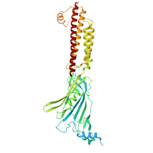
Structure of tetrameric forms of the serotonin-gated 5-HT3 A receptor ion channel.

Introini, B.Cui, W.Chu, X.Zhang, Y.Alves, A.C.Eckhardt-Strelau, L.Golusik, S.Tol, M.Vogel, H.Yuan, S.Kudryashev, M.

(2024) EMBO J 43: 4451-4471

  • DOI: https://doi.org/10.1038/s44318-024-00191-5
  • Primary Citation of Related Structures:  
    8C1W, 8C1Z, 8C20, 8C21

  • PubMed Abstract: 

    Multimeric membrane proteins are produced in the endoplasmic reticulum and transported to their target membranes which, for ion channels, is typically the plasma membrane. Despite the availability of many fully assembled channel structures, our understanding of assembly intermediates, multimer assembly mechanisms, and potential functions of non-standard assemblies is limited. We demonstrate that the pentameric ligand-gated serotonin 5-HT3A receptor (5-HT3AR) can assemble to tetrameric forms and report the structures of the tetramers in plasma membranes of cell-derived microvesicles and in membrane memetics using cryo-electron microscopy and tomography. The tetrameric structures have near-symmetric transmembrane domains, and asymmetric extracellular domains, and can bind serotonin molecules. Computer simulations, based on our cryo-EM structures, were used to decipher the assembly pathway of pentameric 5-HT3R and suggest a potential functional role for the tetrameric receptors.


  • Organizational Affiliation

    Max Planck Institute of Biophysics, Frankfurt am Main, Germany.


Macromolecules
Find similar proteins by:  (by identity cutoff)  |  3D Structure
Entity ID: 1
MoleculeChains Sequence LengthOrganismDetailsImage
5-hydroxytryptamine receptor 3A
A, B, C, D
538Mus musculusMutation(s): 1 
Gene Names: Htr3a5ht3Htr3
Membrane Entity: Yes 
UniProt & NIH Common Fund Data Resources
Find proteins for P23979 (Mus musculus)
Explore P23979 
Go to UniProtKB:  P23979
IMPC:  MGI:96282
Entity Groups  
Sequence Clusters30% Identity50% Identity70% Identity90% Identity95% Identity100% Identity
UniProt GroupP23979
Sequence Annotations
Expand
  • Reference Sequence
Small Molecules
Binding Affinity Annotations 
IDSourceBinding Affinity
SRO BindingDB:  8C21 Ki: min: 66, max: 390 (nM) from 8 assay(s)
IC50: 727 (nM) from 1 assay(s)
EC50: min: 141, max: 1.20e+4 (nM) from 5 assay(s)
Experimental Data & Validation

Experimental Data

  • Method: ELECTRON MICROSCOPY
  • Resolution: 3.60 Å
  • Aggregation State: PARTICLE 
  • Reconstruction Method: SINGLE PARTICLE 
EM Software:
TaskSoftware PackageVersion
MODEL REFINEMENTPHENIX

Structure Validation

View Full Validation Report


Entry History & Funding Information

Deposition Data


Funding OrganizationLocationGrant Number
Alexander von Humboldt FoundationGermanySofja Kovalevskaja Award to MK

Revision History  (Full details and data files)

  • Version 1.0: 2024-07-10
    Type: Initial release
  • Version 1.1: 2024-10-23
    Changes: Data collection, Database references, Structure summary