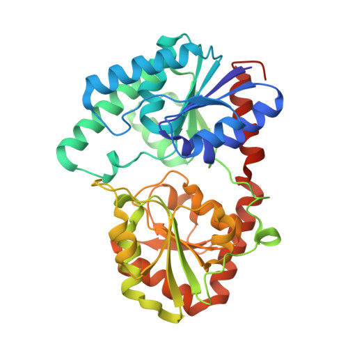
Specificity determinants revealed by the structure of glycosyltransferase Campylobacter concisus PglA.
Vuksanovic, N., Clasman, J.R., Imperiali, B., Allen, K.N.(2024) Protein Sci 33: e4848-e4848
- PubMed: 38019455
- DOI: https://doi.org/10.1002/pro.4848
- Primary Citation of Related Structures:
8DQD, 8DVW, 8DVZ - PubMed Abstract:
In selected Campylobacter species, the biosynthesis of N-linked glycoconjugates via the pgl pathway is essential for pathogenicity and survival. However, most of the membrane-associated GT-B fold glycosyltransferases responsible for diversifying glycans in this pathway have not been structurally characterized which hinders the understanding of the structural factors that govern substrate specificity and prediction of resulting glycan composition. Herein, we report the 1.8 Å resolution structure of Campylobacter concisus PglA, the glycosyltransferase responsible for the transfer of N-acetylgalatosamine (GalNAc) from uridine 5'-diphospho-N-acetylgalactosamine (UDP-GalNAc) to undecaprenyl-diphospho-N,N'-diacetylbacillosamine (UndPP-diNAcBac) in complex with the sugar donor GalNAc. This study identifies distinguishing characteristics that set PglA apart within the GT4 enzyme family. Computational docking of the structure in the membrane in comparison to homologs points to differences in interactions with the membrane-embedded acceptor and the structural analysis of the complex together with bioinformatics and site-directed mutagenesis identifies donor sugar binding motifs. Notably, E113, conserved solely among PglA enzymes, forms a hydrogen bond with the GalNAc C6″-OH. Mutagenesis of E113 reveals activity consistent with this role in substrate binding, rather than stabilization of the oxocarbenium ion transition state, a function sometimes ascribed to the corresponding residue in GT4 homologs. The bioinformatic analyses reveal a substrate-specificity motif, showing that Pro281 in a substrate binding loop of PglA directs configurational preference for GalNAc over GlcNAc. This proline is replaced by a conformationally flexible glycine, even in distant homologs, which favor substrates with the same stereochemistry at C4, such as glucose. The signature loop is conserved across all Campylobacter PglA enzymes, emphasizing its importance in substrate specificity.
Organizational Affiliation:
Department of Chemistry, Boston University, Boston, Massachusetts, USA.


