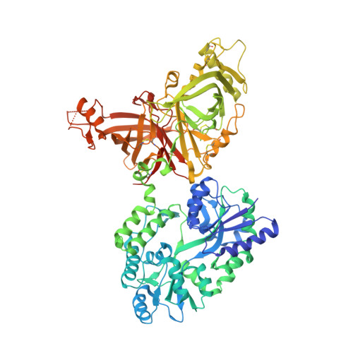
8E0P

Crystal structure of mouse APCDD1 in fusion with engineered MBP


Experimental Data Snapshot

  • Method: X-RAY DIFFRACTION
  • Resolution: 2.33 Å
  • R-Value Free: 0.239 
  • R-Value Work: 0.192 
  • R-Value Observed: 0.193 

Starting Model: experimental
View more details

wwPDB Validation   3D Report Full Report


Ligand Structure Quality Assessment 


This is version 1.2 of the entry. See complete history


Literature

Structure of WNT inhibitor adenomatosis polyposis coli down-regulated 1 (APCDD1), a cell-surface lipid-binding protein.

Hsieh, F.L.Chang, T.H.Gabelli, S.B.Nathans, J.

(2023) Proc Natl Acad Sci U S A 120: e2217096120-e2217096120

  • DOI: https://doi.org/10.1073/pnas.2217096120
  • Primary Citation of Related Structures:  
    8E0P, 8E0R, 8E0W

  • PubMed Abstract: 

    Diverse extracellular proteins negatively regulate WNT signaling. One such regulator is adenomatosis polyposis coli down-regulated 1 (APCDD1), a conserved single-span transmembrane protein. In response to WNT signaling in a variety of tissues, APCDD1 transcripts are highly up-regulated. We have determined the three-dimensional structure of the extracellular domain of APCDD1, and this structure reveals an unusual architecture consisting of two closely apposed β-barrel domains (ABD1 and ABD2). ABD2, but not ABD1, has a large hydrophobic pocket that accommodates a bound lipid. The APCDD1 ECD can also bind to WNT7A, presumably via its covalently bound palmitoleate, a modification that is common to all WNTs and is essential for signaling. This work suggests that APCDD1 functions as a negative feedback regulator by titrating WNT ligands at the surface of responding cells.


  • Organizational Affiliation

    Department of Molecular Biology and Genetics, Johns Hopkins University School of Medicine, Baltimore, MD 21205.


Macromolecules
Find similar proteins by:  (by identity cutoff)  |  3D Structure
Entity ID: 1
MoleculeChains Sequence LengthOrganismDetailsImage
Maltodextrin-binding protein, Protein APCDD1 complex
A, B, C, D
818Escherichia coliMus musculus
This entity is chimeric
Mutation(s): 12 
UniProt & NIH Common Fund Data Resources
Find proteins for C3SHQ8 (Escherichia coli)
Explore C3SHQ8 
Go to UniProtKB:  C3SHQ8
Find proteins for Q3U128 (Mus musculus)
Explore Q3U128 
Go to UniProtKB:  Q3U128
IMPC:  MGI:3513977
Entity Groups  
Sequence Clusters30% Identity50% Identity70% Identity90% Identity95% Identity100% Identity
UniProt GroupsC3SHQ8Q3U128
Glycosylation
Glycosylation Sites: 3Go to GlyGen: Q3U128-1
Sequence Annotations
Expand
  • Reference Sequence
Oligosaccharides

Help

Entity ID: 2
MoleculeChains Length2D Diagram Glycosylation3D Interactions
alpha-D-glucopyranose-(1-4)-beta-D-glucopyranoseE [auth F],
F [auth G],
G [auth H],
H [auth I]
2N/A
Glycosylation Resources
GlyTouCan:  G66120GK
GlyCosmos:  G66120GK
Small Molecules
Ligands 5 Unique
IDChains Name / Formula / InChI Key2D Diagram3D Interactions
DMX
Query on DMX

Download Ideal Coordinates CCD File 
FA [auth D],
Y [auth C],
Z [auth C]
3-[BENZYL(DIMETHYL)AMMONIO]PROPANE-1-SULFONATE
C12 H19 N O3 S
MEJASPJNLSQOAG-UHFFFAOYSA-N
PLM (Subject of Investigation/LOI)
Query on PLM

Download Ideal Coordinates CCD File 
AA [auth C],
L [auth A]
PALMITIC ACID
C16 H32 O2
IPCSVZSSVZVIGE-UHFFFAOYSA-N
NAG
Query on NAG

Download Ideal Coordinates CCD File 
BA [auth D]
CA [auth D]
DA [auth D]
I [auth A]
J [auth A]
BA [auth D],
CA [auth D],
DA [auth D],
I [auth A],
J [auth A],
K [auth A],
O [auth B],
P [auth B],
Q [auth B],
U [auth C],
V [auth C],
W [auth C]
2-acetamido-2-deoxy-beta-D-glucopyranose
C8 H15 N O6
OVRNDRQMDRJTHS-FMDGEEDCSA-N
GOL
Query on GOL

Download Ideal Coordinates CCD File 
EA [auth D],
M [auth A],
R [auth B],
S [auth B],
X [auth C]
GLYCEROL
C3 H8 O3
PEDCQBHIVMGVHV-UHFFFAOYSA-N
CL
Query on CL

Download Ideal Coordinates CCD File 
GA [auth D],
N [auth A],
T [auth B]
CHLORIDE ION
Cl
VEXZGXHMUGYJMC-UHFFFAOYSA-M
Experimental Data & Validation

Experimental Data

  • Method: X-RAY DIFFRACTION
  • Resolution: 2.33 Å
  • R-Value Free: 0.239 
  • R-Value Work: 0.192 
  • R-Value Observed: 0.193 
  • Space Group: P 1 21 1
Unit Cell:
Length ( Å )Angle ( ˚ )
a = 123.914α = 90
b = 98.361β = 109.73
c = 156.513γ = 90
Software Package:
Software NamePurpose
PHENIXrefinement
PHASERphasing
PDB_EXTRACTdata extraction
xia2data reduction
STARANISOdata scaling

Structure Validation

View Full Validation Report


Ligand Structure Quality Assessment 


Entry History & Funding Information

Deposition Data


Funding OrganizationLocationGrant Number
National Institutes of Health/National Eye Institute (NIH/NEI)United States--
Howard Hughes Medical Institute (HHMI)United States--

Revision History  (Full details and data files)

  • Version 1.0: 2023-05-03
    Type: Initial release
  • Version 1.1: 2023-05-24
    Changes: Database references
  • Version 1.2: 2023-10-25
    Changes: Data collection, Refinement description