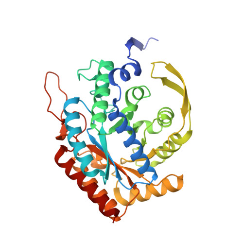
8E0V

DAHP (3-deoxy-D-arabinoheptulosonate-7-phosphate) Synthase complexed with Mn(II), PEP, and Pi in unbound:(bound)2:other Conformations


Experimental Data Snapshot

  • Method: X-RAY DIFFRACTION
  • Resolution: 2.30 Å
  • R-Value Free: 0.228 
  • R-Value Work: 0.194 
  • R-Value Observed: 0.195 

Starting Model: experimental
View more details

wwPDB Validation   3D Report Full Report


Ligand Structure Quality Assessment 


This is version 1.2 of the entry. See complete history


Literature

Role of Half-of-Sites Reactivity and Inter-Subunit Communications in DAHP Synthase Catalysis and Regulation.

Balachandran, N.Grainger, R.A.Rob, T.Liuni, P.Wilson, D.J.Junop, M.S.Berti, P.J.

(2022) Biochemistry 61: 2229-2240

  • DOI: https://doi.org/10.1021/acs.biochem.2c00465
  • Primary Citation of Related Structures:  
    8E0S, 8E0T, 8E0U, 8E0V, 8E0X, 8E0Y, 8E0Z

  • PubMed Abstract: 

    α-Carboxyketose synthases, including 3-deoxy-d- arabino heptulosonate 7-phosphate synthase (DAHPS), are long-standing targets for inhibition. They are challenging targets to create tight-binding inhibitors against, and inhibitors often display half-of-sites binding and partial inhibition. Half-of-sites inhibition demonstrates the existence of inter-subunit communication in DAHPS. We used X-ray crystallography and spatially resolved hydrogen-deuterium exchange (HDX) to reveal the structural and dynamic bases for inter-subunit communication in Escherichia coli DAHPS(Phe), the isozyme that is feedback-inhibited by phenylalanine. Crystal structures of this homotetrameric (dimer-of-dimers) enzyme are invariant over 91% of its sequence. Three variable loops make up 8% of the sequence and are all involved in inter-subunit contacts across the tight-dimer interface. The structures have pseudo-twofold symmetry indicative of inter-subunit communication across the loose-dimer interface, with the diagonal subunits B and C always having the same conformation as each other, while subunits A and D are variable. Spatially resolved HDX reveals contrasting responses to ligand binding, which, in turn, affect binding of the second substrate, erythrose-4-phosphate (E4P). The N -terminal peptide, M1-E12, and the active site loop that binds E4P, F95-K105, are key parts of the communication network. Inter-subunit communication appears to have a catalytic role in all α-carboxyketose synthase families and a regulatory role in some members.


  • Organizational Affiliation

    Department of Biochemistry, Molecular Biology Lab, Western University, London, Ontario N6A 5C1, Canada.


Macromolecules
Find similar proteins by:  (by identity cutoff)  |  3D Structure
Entity ID: 1
MoleculeChains Sequence LengthOrganismDetailsImage
Phospho-2-dehydro-3-deoxyheptonate aldolase, Phe-sensitive
A, B, C, D
351Escherichia coli K-12Mutation(s): 0 
Gene Names: aroGb0754JW0737
EC: 2.5.1.54
UniProt
Find proteins for P0AB91 (Escherichia coli (strain K12))
Explore P0AB91 
Go to UniProtKB:  P0AB91
Entity Groups  
Sequence Clusters30% Identity50% Identity70% Identity90% Identity95% Identity100% Identity
UniProt GroupP0AB91
Sequence Annotations
Expand
  • Reference Sequence
Small Molecules
Ligands 4 Unique
IDChains Name / Formula / InChI Key2D Diagram3D Interactions
GAL
Query on GAL

Download Ideal Coordinates CCD File 
F [auth A],
G [auth A],
N [auth C]
beta-D-galactopyranose
C6 H12 O6
WQZGKKKJIJFFOK-FPRJBGLDSA-N
PEP (Subject of Investigation/LOI)
Query on PEP

Download Ideal Coordinates CCD File 
I [auth B],
L [auth C]
PHOSPHOENOLPYRUVATE
C3 H5 O6 P
DTBNBXWJWCWCIK-UHFFFAOYSA-N
PO4 (Subject of Investigation/LOI)
Query on PO4

Download Ideal Coordinates CCD File 
J [auth B],
M [auth C]
PHOSPHATE ION
O4 P
NBIIXXVUZAFLBC-UHFFFAOYSA-K
MN (Subject of Investigation/LOI)
Query on MN

Download Ideal Coordinates CCD File 
E [auth A],
H [auth B],
K [auth C],
O [auth D]
MANGANESE (II) ION
Mn
WAEMQWOKJMHJLA-UHFFFAOYSA-N
Experimental Data & Validation

Experimental Data

  • Method: X-RAY DIFFRACTION
  • Resolution: 2.30 Å
  • R-Value Free: 0.228 
  • R-Value Work: 0.194 
  • R-Value Observed: 0.195 
  • Space Group: C 1 2 1
Unit Cell:
Length ( Å )Angle ( ˚ )
a = 210.193α = 90
b = 52.908β = 115.711
c = 150.481γ = 90
Software Package:
Software NamePurpose
PHENIXrefinement
autoPROCdata reduction
autoPROCdata scaling
PHASERphasing

Structure Validation

View Full Validation Report


Ligand Structure Quality Assessment 


Entry History & Funding Information

Deposition Data


Funding OrganizationLocationGrant Number
Natural Sciences and Engineering Research Council (NSERC, Canada)Canada2017-06712
Natural Sciences and Engineering Research Council (NSERC, Canada)Canada262034-2013
Natural Sciences and Engineering Research Council (NSERC, Canada)CanadaRGPIN 480432
Canadian Institutes of Health Research (CIHR)CanadaMOP-64422
Canadian Institutes of Health Research (CIHR)CanadaMOP-166070

Revision History  (Full details and data files)

  • Version 1.0: 2022-10-12
    Type: Initial release
  • Version 1.1: 2022-11-02
    Changes: Database references
  • Version 1.2: 2023-10-18
    Changes: Data collection, Refinement description