Targeted covalent inhibitors of the small GTPase Rab27A
De Vita, E., Brustur, D., Tersa, M., Petracca, R., Morgan, R.M.L., Lanyon-Hogg, T., Norman, J.C., Cota, E., Tate, E.W.To be published.
Experimental Data Snapshot
Starting Model: experimental
View more details
Entity ID: 1 | |||||
|---|---|---|---|---|---|
| Molecule | Chains | Sequence Length | Organism | Details | Image |
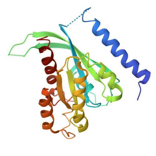
| Synaptotagmin-like protein 2,Ras-related protein Rab-27A | 230 | Homo sapiens | Mutation(s): 2 Gene Names: SYTL2, KIAA1597, SGA72M, SLP2, SLP2A, RAB27A, RAB27 EC: 3.6.5.2 |  | |
UniProt & NIH Common Fund Data Resources | |||||
Find proteins for P51159 (Homo sapiens) Explore P51159 Go to UniProtKB: P51159 | |||||
PHAROS: P51159 GTEx: ENSG00000069974 | |||||
Find proteins for Q9HCH5 (Homo sapiens) Explore Q9HCH5 Go to UniProtKB: Q9HCH5 | |||||
PHAROS: Q9HCH5 GTEx: ENSG00000137501 | |||||
Entity Groups | |||||
| Sequence Clusters | 30% Identity50% Identity70% Identity90% Identity95% Identity100% Identity | ||||
| UniProt Groups | P51159Q9HCH5 | ||||
Sequence AnnotationsExpand | |||||
| |||||
| Ligands 4 Unique | |||||
|---|---|---|---|---|---|
| ID | Chains | Name / Formula / InChI Key | 2D Diagram | 3D Interactions | |
| GNP Query on GNP | C [auth A], I [auth B] | PHOSPHOAMINOPHOSPHONIC ACID-GUANYLATE ESTER C10 H17 N6 O13 P3 UQABYHGXWYXDTK-UUOKFMHZSA-N |  | ||
| WTO (Subject of Investigation/LOI) Query on WTO | G [auth A], K [auth B] | methyl (2~{S})-1-[4-(propanoylamino)phenyl]sulfonylpyrrolidine-2-carboxylate C15 H20 N2 O5 S XUWWNCVLMRGMIO-ZDUSSCGKSA-N |  | ||
| GOL Query on GOL | D [auth A], E [auth A], F [auth A], J [auth B] | GLYCEROL C3 H8 O3 PEDCQBHIVMGVHV-UHFFFAOYSA-N |  | ||
| MG Query on MG | H [auth A], L [auth B] | MAGNESIUM ION Mg JLVVSXFLKOJNIY-UHFFFAOYSA-N |  | ||
| Length ( Å ) | Angle ( ˚ ) |
|---|---|
| a = 60.172 | α = 90 |
| b = 77.696 | β = 90 |
| c = 116.15 | γ = 90 |
| Software Name | Purpose |
|---|---|
| PHENIX | refinement |
| xia2 | data reduction |
| DIALS | data scaling |
| PHASER | phasing |
| Funding Organization | Location | Grant Number |
|---|---|---|
| Cancer Research UK | United Kingdom | C29637/A20781 |