8QCE | pdb_00008qce

Dispersin from Lactiplantibacillus paraplantarum DispLp


Experimental Data Snapshot

  • Method: X-RAY DIFFRACTION
  • Resolution: 1.05 Å
  • R-Value Free: 
    0.170 (Depositor), 0.170 (DCC) 
  • R-Value Work: 
    0.147 (Depositor), 0.147 (DCC) 

Starting Model: experimental
View more details

wwPDB Validation   3D Report Full Report


Ligand Structure Quality Assessment 

Created with Raphaël 2.3.0Worse 01 BetterLigand structure goodness of fit to experimental dataBest fitted GC2Click on this verticalbar to view details

This is version 1.1 of the entry. See complete history


Literature

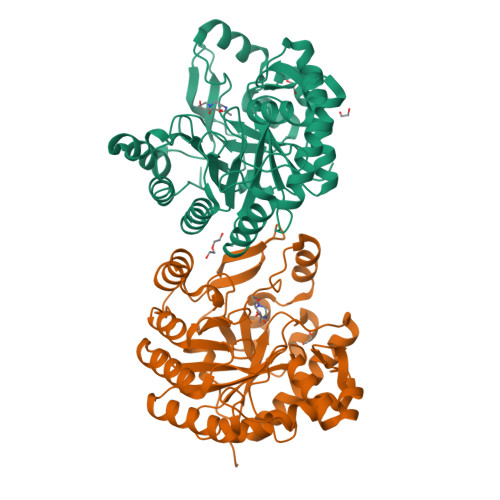
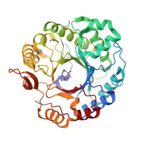
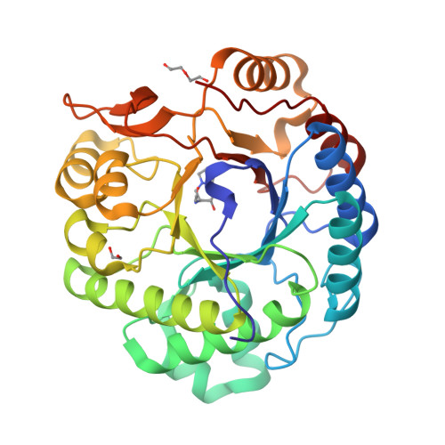
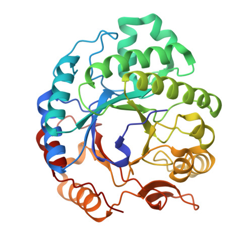
Expansion of the diversity of dispersin scaffolds.

Males, A.Moroz, O.V.Blagova, E.Munch, A.Hansen, G.H.Johansen, A.H.Ostergaard, L.H.Segura, D.R.Eddenden, A.Due, A.V.Gudmand, M.Salomon, J.Sorensen, S.R.Franco Cairo, J.P.L.Nitz, M.Pache, R.A.Vejborg, R.M.Bhosale, S.Vocadlo, D.J.Davies, G.J.Wilson, K.S.

(2025) Acta Crystallogr D Struct Biol 81: 130-146

  • DOI: https://doi.org/10.1107/S205979832500110X
  • Primary Citation of Related Structures:  
    8QAK, 8QB6, 8QCE, 9HTA

  • PubMed Abstract: 

    Microorganisms are known to secrete copious amounts of extracellular polymeric substances (EPS) that form complex matrices around the cells to shield them against external stresses, to maintain structural integrity and to influence their environment. Many microorganisms also secrete enzymes that are capable of remodelling or degrading EPS in response to various environmental cues. One key enzyme class is the poly-β-1,6-linked N-acetyl-D-glucosamine (PNAG)-degrading glycoside hydrolases, of which the canonical member is dispersin B (DspB) from CAZy family GH20. We sought to test the hypothesis that PNAG-degrading enzymes would be present across family GH20, resulting in expansion of the sequence and structural space and thus the availability of PNAGases. Phylogenetic analysis revealed that several microorganisms contain potential DspB-like enzymes. Six of these were expressed and characterized, and four crystal structures were determined (two of which were in complex with the established GH20 inhibitor 6-acetamido-6-deoxy-castanospermine and one with a bespoke disaccharide β-1,6-linked thiazoline inhibitor). One enzyme expressed rather poorly, which restricted crystal screening and did not allow activity measurements. Using synthetic PNAG oligomers and MALDI-TOF analysis, two of the five enzymes tested showed preferential endo hydrolytic activity. Their sequences, having only 26% identity to the pioneer enzyme DspB, highlight the considerable array of previously unconsidered dispersins in nature, greatly expanding the range of potential dispersin backbones available for societal application and engineering.


  • Organizational Affiliation

    York Structural Biology Laboratory, Department of Chemistry, University of York, York YO10 5DD, United Kingdom.


Macromolecules
Find similar proteins by:  (by identity cutoff)  |  3D Structure
Entity ID: 1
MoleculeChains Sequence LengthOrganismDetailsImage
N-acetyl-beta-hexosaminidase
A, B
336Lactiplantibacillus paraplantarumMutation(s): 0 
Gene Names: DQM08_04690
Entity Groups  
Sequence Clusters30% Identity50% Identity70% Identity90% Identity95% Identity100% Identity
Sequence Annotations
Expand
  • Reference Sequence
Small Molecules
Ligands 3 Unique
IDChains Name / Formula / InChI Key2D Diagram3D Interactions
GC2 (Subject of Investigation/LOI)
Query on GC2

Download Ideal Coordinates CCD File 
C [auth A],
F [auth B]
6-ACETAMIDO-6-DEOXY-CASTANOSPERMINE
C10 H18 N2 O4
IHKWXDCSAKJQKM-SRQGCSHVSA-N
PEG
Query on PEG

Download Ideal Coordinates CCD File 
H [auth B]DI(HYDROXYETHYL)ETHER
C4 H10 O3
MTHSVFCYNBDYFN-UHFFFAOYSA-N
EDO
Query on EDO

Download Ideal Coordinates CCD File 
D [auth A],
E [auth A],
G [auth B]
1,2-ETHANEDIOL
C2 H6 O2
LYCAIKOWRPUZTN-UHFFFAOYSA-N
Experimental Data & Validation

Experimental Data

  • Method: X-RAY DIFFRACTION
  • Resolution: 1.05 Å
  • R-Value Free:  0.170 (Depositor), 0.170 (DCC) 
  • R-Value Work:  0.147 (Depositor), 0.147 (DCC) 
Space Group: P 1 21 1
Unit Cell:
Length ( Å )Angle ( ˚ )
a = 46.902α = 90
b = 82.792β = 98.115
c = 80.642γ = 90
Software Package:
Software NamePurpose
REFMACrefinement
REFMACrefinement
Aimlessdata scaling
XDSdata reduction
Aimlessdata scaling
MOLREPphasing

Structure Validation

View Full Validation Report


Ligand Structure Quality Assessment 

Created with Raphaël 2.3.0Worse 01 BetterLigand structure goodness of fit to experimental dataBest fitted GC2Click on this verticalbar to view details

Entry History & Funding Information

Deposition Data


Funding OrganizationLocationGrant Number
Not funded--

Revision History  (Full details and data files)

  • Version 1.0: 2025-03-12
    Type: Initial release
  • Version 1.1: 2025-03-19
    Changes: Database references