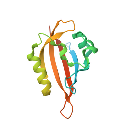
8QIV

CrPhotLOV1 light state structure 87.5 ms (85-90 ms) after illumination determined by time-resolved serial synchrotron crystallography at room temperature


Experimental Data Snapshot

  • Method: X-RAY DIFFRACTION
  • Resolution: 3.10 Å
  • R-Value Free: 0.349 
  • R-Value Work: 0.237 
  • R-Value Observed: 0.243 

Starting Model: experimental
View more details

wwPDB Validation   3D Report Full Report


This is version 1.3 of the entry. See complete history


Literature

Capturing the blue-light activated state of the Phot-LOV1 domain from Chlamydomonas reinhardtii using time-resolved serial synchrotron crystallography.

Gotthard, G.Mous, S.Weinert, T.Maia, R.N.A.James, D.Dworkowski, F.Gashi, D.Furrer, A.Ozerov, D.Panepucci, E.Wang, M.Schertler, G.F.X.Heberle, J.Standfuss, J.Nogly, P.

(2024) IUCrJ 11: 792-808

  • DOI: https://doi.org/10.1107/S2052252524005608
  • Primary Citation of Related Structures:  
    8QI8, 8QI9, 8QIA, 8QIB, 8QIF, 8QIG, 8QIH, 8QII, 8QIK, 8QIL, 8QIM, 8QIN, 8QIO, 8QIP, 8QIQ, 8QIR, 8QIS, 8QIT, 8QIU, 8QIV, 8QIW

  • PubMed Abstract: 

    Light-oxygen-voltage (LOV) domains are small photosensory flavoprotein modules that allow the conversion of external stimuli (sunlight) into intracellular signals responsible for various cell behaviors (e.g. phototropism and chloroplast relocation). This ability relies on the light-induced formation of a covalent thioether adduct between a flavin chromophore and a reactive cysteine from the protein environment, which triggers a cascade of structural changes that result in the activation of a serine/threonine (Ser/Thr) kinase. Recent developments in time-resolved crystallography may allow the activation cascade of the LOV domain to be observed in real time, which has been elusive. In this study, we report a robust protocol for the production and stable delivery of microcrystals of the LOV domain of phototropin Phot-1 from Chlamydomonas reinhardtii (CrPhotLOV1) with a high-viscosity injector for time-resolved serial synchrotron crystallography (TR-SSX). The detailed process covers all aspects, from sample optimization to data collection, which may serve as a guide for soluble protein preparation for TR-SSX. In addition, we show that the crystals obtained preserve the photoreactivity using infrared spectroscopy. Furthermore, the results of the TR-SSX experiment provide high-resolution insights into structural alterations of CrPhotLOV1 from Δt = 2.5 ms up to Δt = 95 ms post-photoactivation, including resolving the geometry of the thioether adduct and the C-terminal region implicated in the signal transduction process.


  • Organizational Affiliation

    Laboratory of Biomolecular Research, Division of Biology and Chemistry, Paul Scherrer Institute, 5232 Villigen PSI, Switzerland.


Macromolecules
Find similar proteins by:  (by identity cutoff)  |  3D Structure
Entity ID: 1
MoleculeChains Sequence LengthOrganismDetailsImage
Phototropin138Chlamydomonas reinhardtiiMutation(s): 0 
Gene Names: PHOTCHLRE_03g199000v5CHLREDRAFT_183965
EC: 2.7.11.1
UniProt
Find proteins for Q8LPD9 (Chlamydomonas reinhardtii)
Explore Q8LPD9 
Go to UniProtKB:  Q8LPD9
Entity Groups  
Sequence Clusters30% Identity50% Identity70% Identity90% Identity95% Identity100% Identity
UniProt GroupQ8LPD9
Sequence Annotations
Expand
  • Reference Sequence
Small Molecules
Ligands 1 Unique
IDChains Name / Formula / InChI Key2D Diagram3D Interactions
FMN (Subject of Investigation/LOI)
Query on FMN

Download Ideal Coordinates CCD File 
B [auth A]FLAVIN MONONUCLEOTIDE
C17 H21 N4 O9 P
FVTCRASFADXXNN-SCRDCRAPSA-N
Experimental Data & Validation

Experimental Data

  • Method: X-RAY DIFFRACTION
  • Resolution: 3.10 Å
  • R-Value Free: 0.349 
  • R-Value Work: 0.237 
  • R-Value Observed: 0.243 
  • Space Group: P 65 2 2
Unit Cell:
Length ( Å )Angle ( ˚ )
a = 121.54α = 90
b = 121.54β = 90
c = 46.18γ = 120
Software Package:
Software NamePurpose
PHENIXrefinement
CrystFELdata reduction
XSCALEdata scaling
PHENIXphasing

Structure Validation

View Full Validation Report


Entry History & Funding Information

Deposition Data


Funding OrganizationLocationGrant Number
H2020 Marie Curie Actions of the European CommissionEuropean Union701647
Swiss National Science FoundationSwitzerlandPZ00P3_174169
Polish National Science CentrePolandUMO-2021/03/H/NZ1/00002
Swiss National Science FoundationSwitzerland192760

Revision History  (Full details and data files)

  • Version 1.0: 2024-07-24
    Type: Initial release
  • Version 1.1: 2024-07-31
    Changes: Database references
  • Version 1.2: 2024-09-11
    Changes: Database references
  • Version 1.3: 2024-11-06
    Changes: Structure summary