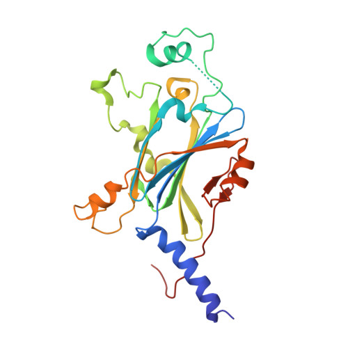
8QQC

Crystal structure of Lymantria dispar CPV14 polyhedra single crystal


Experimental Data Snapshot

  • Method: X-RAY DIFFRACTION
  • Resolution: 1.30 Å
  • R-Value Free: 0.257 
  • R-Value Work: 0.229 
  • R-Value Observed: 0.230 

Starting Model: experimental
View more details

wwPDB Validation   3D Report Full Report


Ligand Structure Quality Assessment 


This is version 1.2 of the entry. See complete history

Re-refinement Note

This entry reflects an alternative modeling of the original data in: 8QPH


Literature

VMXm - A sub-micron focus macromolecular crystallography beamline at Diamond Light Source.

Warren, A.J.Trincao, J.Crawshaw, A.D.Beale, E.V.Duller, G.Stallwood, A.Lunnon, M.Littlewood, R.Prescott, A.Foster, A.Smith, N.Rehm, G.Gayadeen, S.Bloomer, C.Alianelli, L.Laundy, D.Sutter, J.Cahill, L.Evans, G.

(2024) J Synchrotron Radiat 31: 1593-1608

  • DOI: https://doi.org/10.1107/S1600577524009160
  • Primary Citation of Related Structures:  
    8QPH, 8QQC

  • PubMed Abstract: 

    VMXm joins the suite of operational macromolecular crystallography beamlines at Diamond Light Source. It has been designed to optimize rotation data collections from protein crystals less than 10 µm and down to below 1 µm in size. The beamline has a fully focused beam of 0.3 × 2.3 µm (vertical × horizontal) with a tuneable energy range (6-28 keV) and high flux (1.6 × 10 12  photons s -1 at 12.5 keV). The crystals are housed within a vacuum chamber to minimize background scatter from air. Crystals are plunge-cooled on cryo-electron microscopy grids, allowing much of the liquid surrounding the crystals to be removed. These factors improve the signal-to-noise during data collection and the lifetime of the microcrystals can be prolonged by exploiting photoelectron escape. A novel in vacuo sample environment has been designed which also houses a scanning electron microscope to aid with sample visualization. This combination of features at VMXm allows measurements at the physical limits of X-ray crystallography on biomacromolecules to be explored and exploited.


  • Organizational Affiliation

    Diamond Light Source, Harwell Science and Innovation Campus, Didcot, Oxfordshire OX11 0DE, United Kingdom.


Macromolecules
Find similar proteins by:  (by identity cutoff)  |  3D Structure
Entity ID: 1
MoleculeChains Sequence LengthOrganismDetailsImage
Polyhedrin249Lymantria disparMutation(s): 0 
Gene Names: pod
UniProt
Find proteins for Q91IE3 (Lymantria dispar cypovirus 14)
Explore Q91IE3 
Go to UniProtKB:  Q91IE3
Entity Groups  
Sequence Clusters30% Identity50% Identity70% Identity90% Identity95% Identity100% Identity
UniProt GroupQ91IE3
Sequence Annotations
Expand
  • Reference Sequence
Experimental Data & Validation

Experimental Data

  • Method: X-RAY DIFFRACTION
  • Resolution: 1.30 Å
  • R-Value Free: 0.257 
  • R-Value Work: 0.229 
  • R-Value Observed: 0.230 
  • Space Group: I 2 3
Unit Cell:
Length ( Å )Angle ( ˚ )
a = 103.102α = 90
b = 103.102β = 90
c = 103.102γ = 90
Software Package:
Software NamePurpose
PHENIXrefinement
DIALSdata reduction
xia2.multiplexdata scaling
PHASERphasing

Structure Validation

View Full Validation Report


Ligand Structure Quality Assessment 


Entry History & Funding Information

Deposition Data


Funding OrganizationLocationGrant Number
Not funded--

Revision History  (Full details and data files)

  • Version 1.0: 2023-11-08
    Type: Initial release
  • Version 1.1: 2024-11-13
    Changes: Database references, Structure summary
  • Version 1.2: 2024-11-20
    Changes: Database references