8SNI | pdb_00008sni

Hydroxynitrile Lyase from Hevea brasiliensis with Forty Mutations


Experimental Data Snapshot

  • Method: X-RAY DIFFRACTION
  • Resolution: 1.99 Å
  • R-Value Free: 
    0.227 (Depositor), 0.227 (DCC) 
  • R-Value Work: 
    0.199 (Depositor), 0.199 (DCC) 
  • R-Value Observed: 
    0.200 (Depositor) 

Starting Model: experimental
View more details

wwPDB Validation   3D Report Full Report


Ligand Structure Quality Assessment 

Created with Raphaël 2.3.0Worse 01 BetterLigand structure goodness of fit to experimental dataBest fitted PROClick on this verticalbar to view detailsBest fitted SO4Click on this verticalbar to view detailsBest fitted EDOClick on this verticalbar to view details

This is version 2.1 of the entry. See complete history


Literature

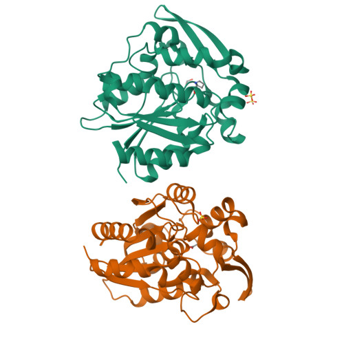
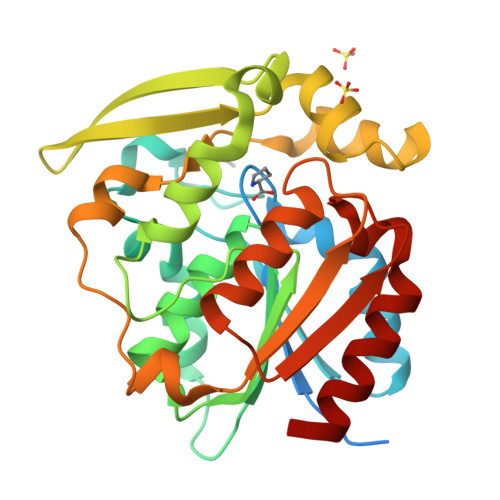
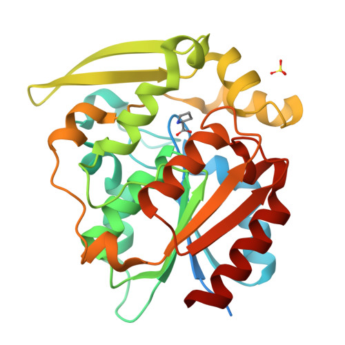
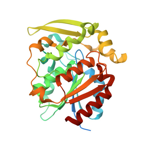
Crystal structures of 40- and 71-substitution variants of hydroxynitrile lyase from rubber tree.

Pierce, C.T.Tan, P.Greenberg, L.R.Walsh, M.E.Shi, K.Nguyen, A.H.Meixner, E.L.Sarak, S.Aihara, H.Evans 3rd, R.L.Kazlauskas, R.J.

(2025) Acta Crystallogr D Struct Biol 81: 511-523

  • DOI: https://doi.org/10.1107/S2059798325007065
  • Primary Citation of Related Structures:  
    8SNI, 9CLR

  • PubMed Abstract: 

    Hydroxynitrile lyase from Hevea brasiliensis (HbHNL) and the esterase SABP2 from Nicotiana tabacum share the α/β-hydrolase fold, a Ser-His-Asp catalytic triad and 44% sequence identity, yet catalyze different reactions. Prior studies showed that three active-site substitutions in HbHNL conferred weak esterase activity. To investigate how regions beyond the active site influence catalytic efficiency and active-site geometry, we engineered HbHNL variants with increasing numbers of substitutions to match SABP2. Variant HNL16 has all amino acids within 6.5 Å of the active site identical to SABP2, HNL40 those within 10 Å and HNL71 those within 14 Å. HNL16 exhibited poor esterase activity, whereas both HNL40 and HNL71 showed efficient esterase catalysis, demonstrating that residues beyond the immediate active site are critical for functional conversion. X-ray structures of HNL40 and HNL71 reveal a progressive shift in backbone positions toward those of SABP2, with r.m.s.d. values of 0.51 Å (HNL40) and 0.41 Å (HNL71) over the C α atoms, and even smaller r.m.s.d.s within the active-site region. Both HNL40 and HNL71 show a restored oxyanion hole and an additional tunnel connecting the active site to the protein surface. This work demonstrates the essential role of distant, indirectly acting residues to catalysis in α/β-hydrolase enzymes.


  • Organizational Affiliation
    • Department of Biochemistry, Molecular Biology and Biophysics, University of Minnesota, Minneapolis, MN 55455, USA.

Macromolecules
Find similar proteins by:  (by identity cutoff)  |  3D Structure
Entity ID: 1
MoleculeChains Sequence LengthOrganismDetailsImage
(S)-hydroxynitrile lyase
A, B
293Hevea brasiliensisMutation(s): 38 
Gene Names: HNL
EC: 4.1.2.47
UniProt
Find proteins for P52704 (Hevea brasiliensis)
Explore P52704 
Go to UniProtKB:  P52704
Entity Groups  
Sequence Clusters30% Identity50% Identity70% Identity90% Identity95% Identity100% Identity
UniProt GroupP52704
Sequence Annotations
Expand
  • Reference Sequence
Small Molecules
Ligands 3 Unique
IDChains Name / Formula / InChI Key2D Diagram3D Interactions
PRO (Subject of Investigation/LOI)
Query on PRO

Download Ideal Coordinates CCD File 
F [auth A],
H [auth B]
PROLINE
C5 H9 N O2
ONIBWKKTOPOVIA-BYPYZUCNSA-N
SO4 (Subject of Investigation/LOI)
Query on SO4

Download Ideal Coordinates CCD File 
D [auth A],
E [auth A],
G [auth B]
SULFATE ION
O4 S
QAOWNCQODCNURD-UHFFFAOYSA-L
EDO (Subject of Investigation/LOI)
Query on EDO

Download Ideal Coordinates CCD File 
C [auth A]1,2-ETHANEDIOL
C2 H6 O2
LYCAIKOWRPUZTN-UHFFFAOYSA-N
Experimental Data & Validation

Experimental Data

  • Method: X-RAY DIFFRACTION
  • Resolution: 1.99 Å
  • R-Value Free:  0.227 (Depositor), 0.227 (DCC) 
  • R-Value Work:  0.199 (Depositor), 0.199 (DCC) 
  • R-Value Observed: 0.200 (Depositor) 
Space Group: P 21 21 21
Unit Cell:
Length ( Å )Angle ( ˚ )
a = 76.527α = 90
b = 81.884β = 90
c = 90.929γ = 90
Software Package:
Software NamePurpose
PHENIXrefinement
XDSdata reduction
XSCALEdata scaling
PHASERphasing

Structure Validation

View Full Validation Report


Ligand Structure Quality Assessment 

Created with Raphaël 2.3.0Worse 01 BetterLigand structure goodness of fit to experimental dataBest fitted PROClick on this verticalbar to view detailsBest fitted SO4Click on this verticalbar to view detailsBest fitted EDOClick on this verticalbar to view details

Entry History & Funding Information

Deposition Data


Funding OrganizationLocationGrant Number
National Institutes of Health/National Institute of General Medical Sciences (NIH/NIGMS)United StatesGM119483
National Institutes of Health/National Institute of General Medical Sciences (NIH/NIGMS)United StatesNIGMS R35-GM118047
National Science Foundation (NSF, United States)United States2039039

Revision History  (Full details and data files)

  • Version 1.0: 2024-02-28
    Type: Initial release
  • Version 2.0: 2025-02-19
    Type: Coordinate replacement
    Reason: Model completeness
    Changes: Advisory, Atomic model, Author supporting evidence, Data collection, Database references, Derived calculations, Non-polymer description, Refinement description, Structure summary
  • Version 2.1: 2025-09-10
    Changes: Database references