8VB0

Asymmetric unit of bacteriophage PhiM1 mature capsid


Experimental Data Snapshot

  • Method: ELECTRON MICROSCOPY
  • Resolution: 3.04 Å
  • Aggregation State: PARTICLE 
  • Reconstruction Method: SINGLE PARTICLE 

wwPDB Validation   3D Report Full Report


This is version 1.0 of the entry. See complete history


Literature

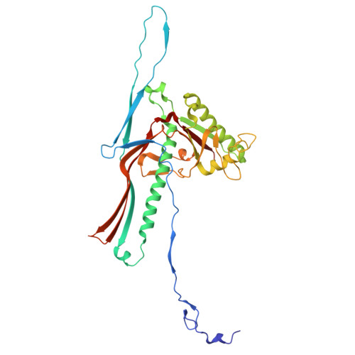
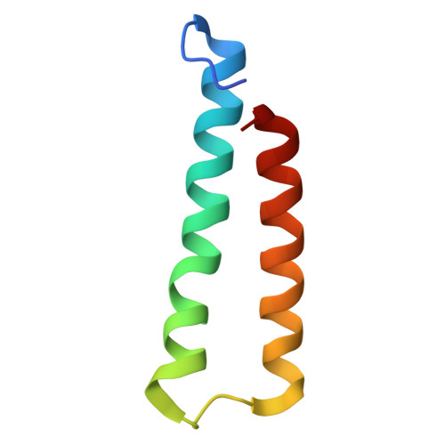
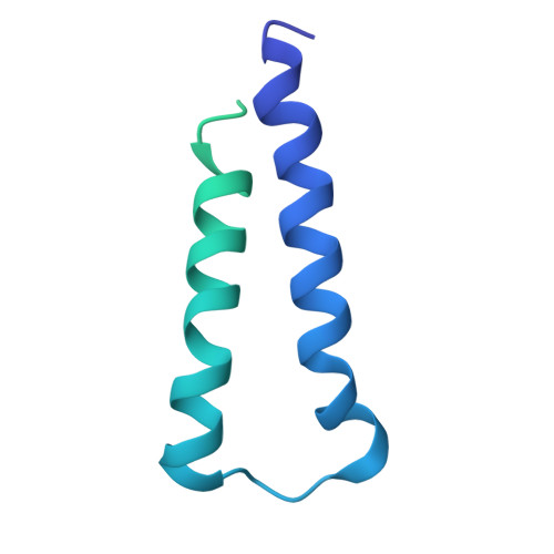
Ejectosome of Pectobacterium bacteriophage Phi M1.

Eruera, A.R.Hodgkinson-Bean, J.Rutter, G.L.Hills, F.R.Kumaran, R.Crowe, A.J.M.Jadav, N.Chang, F.McJarrow-Keller, K.Jorge, F.Hyun, J.Kim, H.Ryu, B.Bostina, M.

(2024) PNAS Nexus 3: pgae416-pgae416

  • DOI: https://doi.org/10.1093/pnasnexus/pgae416
  • Primary Citation of Related Structures:  
    8VB0, 8VB2, 8VB4, 8VBX

  • PubMed Abstract: 

    Podophages that infect gram-negative bacteria, such as Pectobacterium pathogen ΦM1, encode tail assemblies too short to extend across the complex gram-negative cell wall. To overcome this, podophages encode a large protein complex (ejectosome) packaged inside the viral capsid and correspondingly ejected during infection to form a transient channel that spans the periplasmic space. Here, we describe the ejectosome of bacteriophage ΦM1 to a resolution of 3.32 Å by single-particle cryo-electron microscopy (cryo-EM). The core consists of tetrameric and octameric ejection proteins which form a ∼1.5-MDa ejectosome that must transition through the ∼30 Å aperture created by the short tail nozzle assembly that acts as the conduit for the passage of DNA during infection. The ejectosome forms several grooves into which coils of genomic DNA are fit before the DNA sharply turns and goes down the tunnel and into the portal. In addition, we reconstructed the icosahedral capsid and hybrid tail apparatus to resolutions between 3.04 and 3.23 Å, and note an uncommon fold adopted by the dimerized decoration proteins which further emphasize the structural diversity of podophages. These reconstructions have allowed the generation of a complete atomic model of the ΦM1, uncovering two distinct decoration proteins and highlighting the exquisite structural diversity of tailed bacteriophages.


  • Organizational Affiliation

    Department of Microbiology and Immunology, University of Otago, Dunedin 9010, New Zealand.


Macromolecules
Find similar proteins by:  (by identity cutoff)  |  3D Structure
Entity ID: 1
MoleculeChains Sequence LengthOrganismDetailsImage
Major capsid protein (gp38)
A, B, C, D, E
A, B, C, D, E, F, G
327Pectobacterium phage PhiM1Mutation(s): 0 
UniProt
Find proteins for A0A1P7WG08 (Pectobacterium phage PhiM1)
Explore A0A1P7WG08 
Go to UniProtKB:  A0A1P7WG08
Entity Groups  
Sequence Clusters30% Identity50% Identity70% Identity90% Identity95% Identity100% Identity
UniProt GroupA0A1P7WG08
Sequence Annotations
Expand
  • Reference Sequence
Find similar proteins by:  (by identity cutoff)  |  3D Structure
Entity ID: 2
MoleculeChains Sequence LengthOrganismDetailsImage
Alpha-claw decoration protein (gp44)
H, I, K, L, M
62Pectobacterium phage PhiM1Mutation(s): 0 
UniProt
Find proteins for A0A1P7WG15 (Pectobacterium phage PhiM1)
Explore A0A1P7WG15 
Go to UniProtKB:  A0A1P7WG15
Entity Groups  
Sequence Clusters30% Identity50% Identity70% Identity90% Identity95% Identity100% Identity
UniProt GroupA0A1P7WG15
Sequence Annotations
Expand
  • Reference Sequence
Find similar proteins by:  (by identity cutoff)  |  3D Structure
Entity ID: 3
MoleculeChains Sequence LengthOrganismDetailsImage
Alpha-paw decoration protein (gp43)
J, N
179Pectobacterium phage PhiM1Mutation(s): 0 
UniProt
Find proteins for A0A1P7WG13 (Pectobacterium phage PhiM1)
Explore A0A1P7WG13 
Go to UniProtKB:  A0A1P7WG13
Entity Groups  
Sequence Clusters30% Identity50% Identity70% Identity90% Identity95% Identity100% Identity
UniProt GroupA0A1P7WG13
Sequence Annotations
Expand
  • Reference Sequence
Experimental Data & Validation

Experimental Data

  • Method: ELECTRON MICROSCOPY
  • Resolution: 3.04 Å
  • Aggregation State: PARTICLE 
  • Reconstruction Method: SINGLE PARTICLE 

Structure Validation

View Full Validation Report


Entry History & Funding Information

Deposition Data


Funding OrganizationLocationGrant Number
Other privateNew Zealand--

Revision History  (Full details and data files)

  • Version 1.0: 2024-10-23
    Type: Initial release